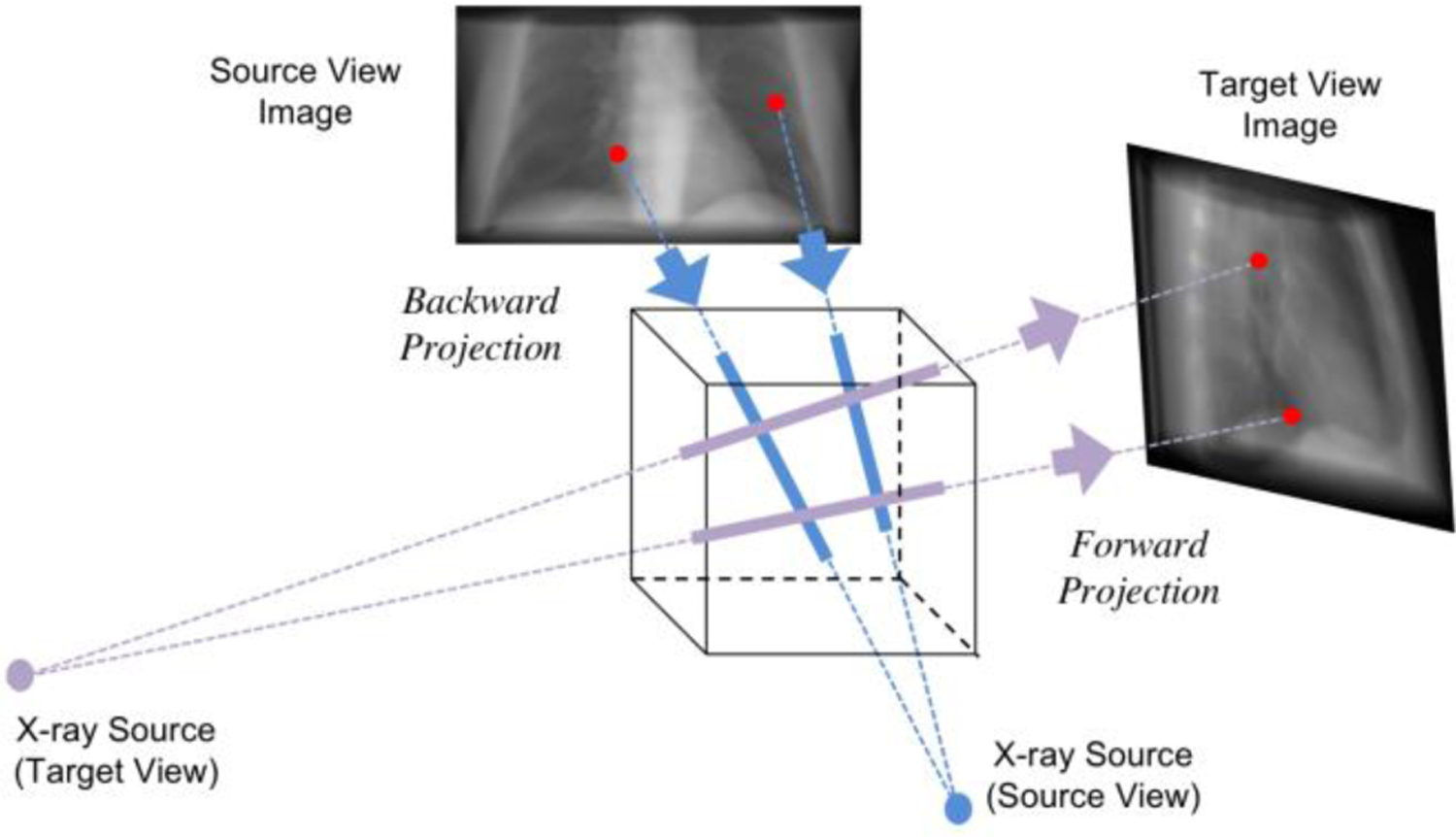
Fig. 3.

Illustration of geometry transformation with backward projection (blue) and forward projection (purple). The back projector puts the pixel intensities in the source-view image back to the corresponding voxels in the 3D volume according to the cone-beam geometry of the physical model. When the X-ray source rotates to the target view angles, the forward projection operator integrates along the projection line and projects onto the detector plane.
