
Fig. 2.
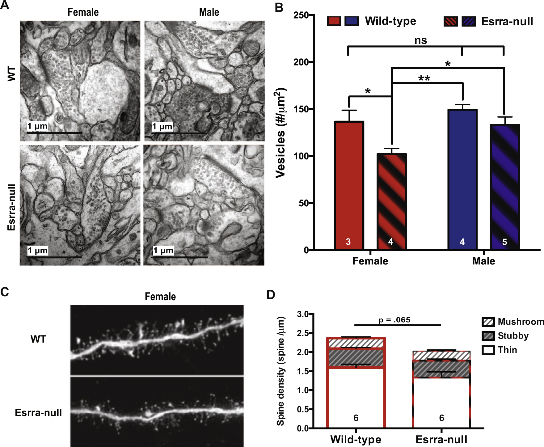
Decreased synaptic vesicle pool density in female Esrra-null mice. (A) Representative electron microscope images from NAc core of both females and male WT and Esrra-null mice. (B) Quantification of vesicle density from 34,535 vesicles in 508 synapses showing a significant reduction in females, but not male, Esrra-null mice. (C) Representative images of medium spiny neurons in the NAc core that were loaded with Lucifer yellow for dendritic spine density assessment in female WT and Esrra-null (KO) mice. (D) Quantification of 15,075 spines shows no statistically significant difference between female WT and Esrra-null mice. Data are presented as mean ± S.E.M. A two-way ANOVA was used in (B) and a Mann–Whitney U test was used for (D). **p < .01, ***p < .001 indicate significant differences between the groups.