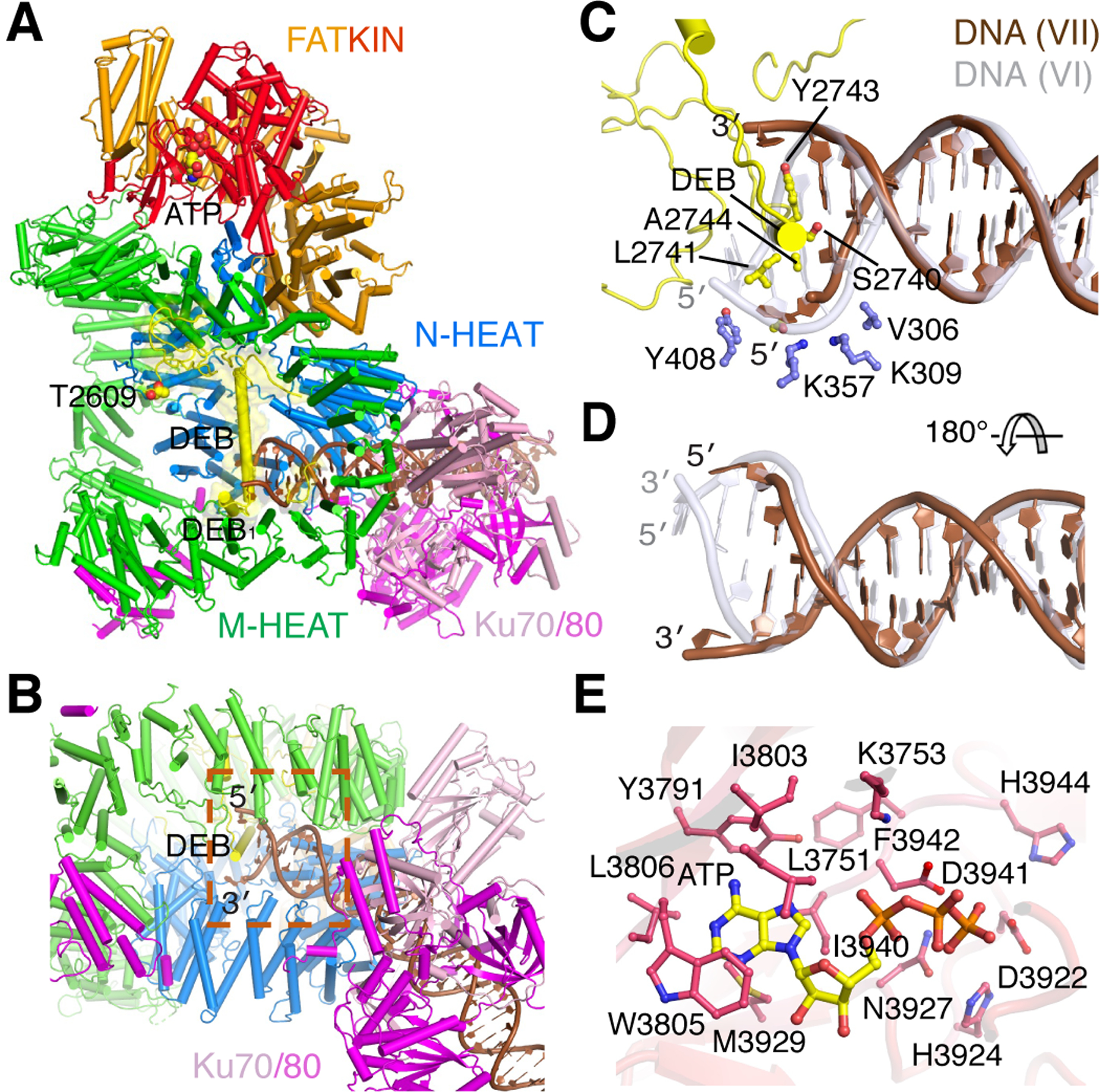
Figure 4. Structure of Complex VII.

A. The overall structure with the unphosphorylated ABCDE patch (yellow) covering the DNA blunt end. B. The local structure of the DNA end sandwiched between two HEAT rings and protected by the long helix DEB of ABCDE. C. Zoom-in view of protein-DNA interactions at the blunt end. The first nucleotide of each strand is unpaired. DNA in Complex VI is shown as semitransparent grey after superposition of DNA-PK. D. The DNA in complex VII is shifted outward by 2 bp along the helical contour. E. The active site of DNA-PKcs is occupied by ATP. The adenine base is cocooned by hydrophobic residues. The catalytic residues are in proximity for phosphorylation of a substrate peptide.
