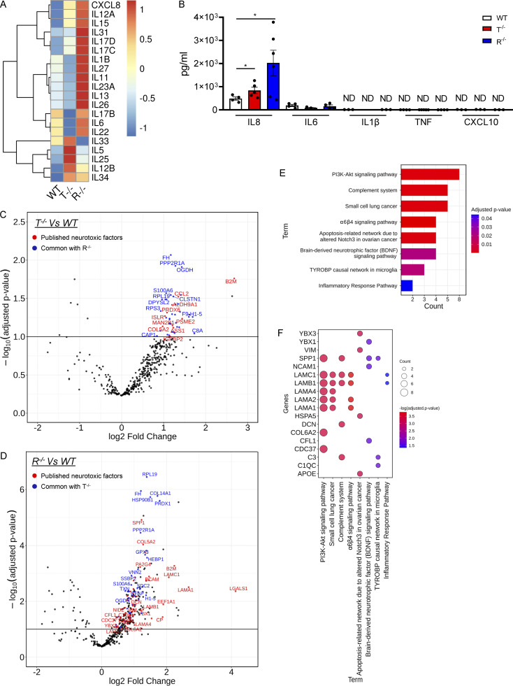
Figure 6.
KO iPSC–derived astrocytes secrete neuroinflammatory mediators. (A) Heatmap representation showing gene expression analysis of interleukin genes in T−/− and R−/− KO cells compared with WT. (B) ELISA results for inflammatory cytokines (IL8, IL6, IL1β, TNFα, and CXCL10) in the supernatant of WT and KO astrocytes at steady state. (Mean ± SEM; n = 3 independent experiments; one-tailed Mann–Whitney U test; *, P < 0.05.) (C and D) Volcano plots highlighting the differentially secreted proteins in R−/− (C) and T−/− (D) compared with WT samples. Log2(FC) and adjusted P values are reported for each protein. Upregulated common secreted proteins between the two comparisons are highlighted in blue. Proteins common with published neurotoxic factors are highlighted in red. (E) Barplot highlighting the significant enriched functional pathways resulting from the WikiPathways database of secreted proteins common with published neurotoxic factor. (F) Bubble plot showing the differentially secreted neurotoxic factors involved in the identified enriched pathways.

