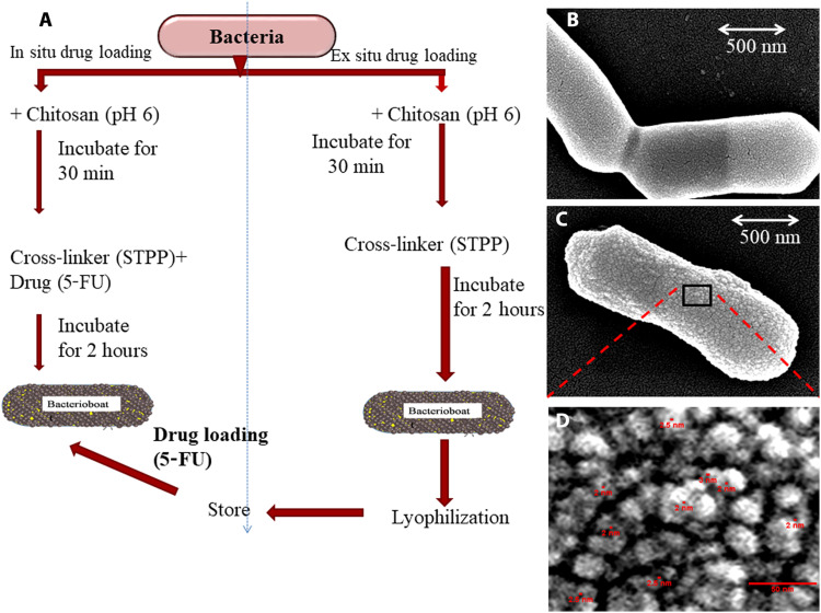
Fig. 1. BB synthesis and morphology.
(A) Schematic representation of the synthesis of BB with in situ and ex situ loading protocol for drugs of choice. (B) FE-SEM image of L. reuteri cells. (C) FE-SEM image of freshly prepared BB showing chitosan nanoparticles on the cell surface. (D) Enlarged view of black box area of (C) illustrating the sponge ball–shaped nanoparticles of 15 to 25 nm in size with a pore diameter of around 2 to 3 nm.

