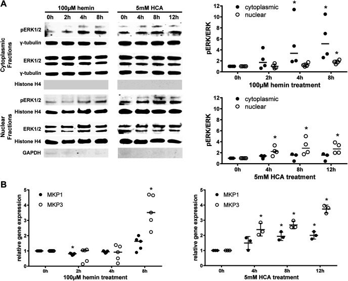
Figure 5.
Hyperactivated ERK1/2 remains in the cytoplasm in hemin-induced ferroptosis and the transcription of Mkp1 and Mkp3, its negative regulators, is delayed. A, The time course of the levels of phospho-ERK and total ERK was assessed in the cytoplasmic and nuclear extracts of primary neurons exposed to 100 μm hemin or 5 mm HCA (normalized to γ-tubulin for cytoplasmic fractions or Histone H4 for nuclear fractions). The fractionation was confirmed by evaluating the Histone H4 expression in the cytoplasmic fractions and the GAPDH expression in the nuclear fractions. Medians are given, except for the nuclear fractions in hemin that are means ± SD; *p < 0.05 versus 0 h. For exact p values, refer to Extended Data Figure 5-1. B, The Mkp1 and Mkp3 mRNA expression was measured in primary neurons exposed to 100 μm hemin or 5 mm HCA. The values represent the medians for hemin and the means ± SD for HCA; *p < 0.05 versus 0 h. For exact p values, refer to Extended Data Figure 5-2.

