
Figure 9.
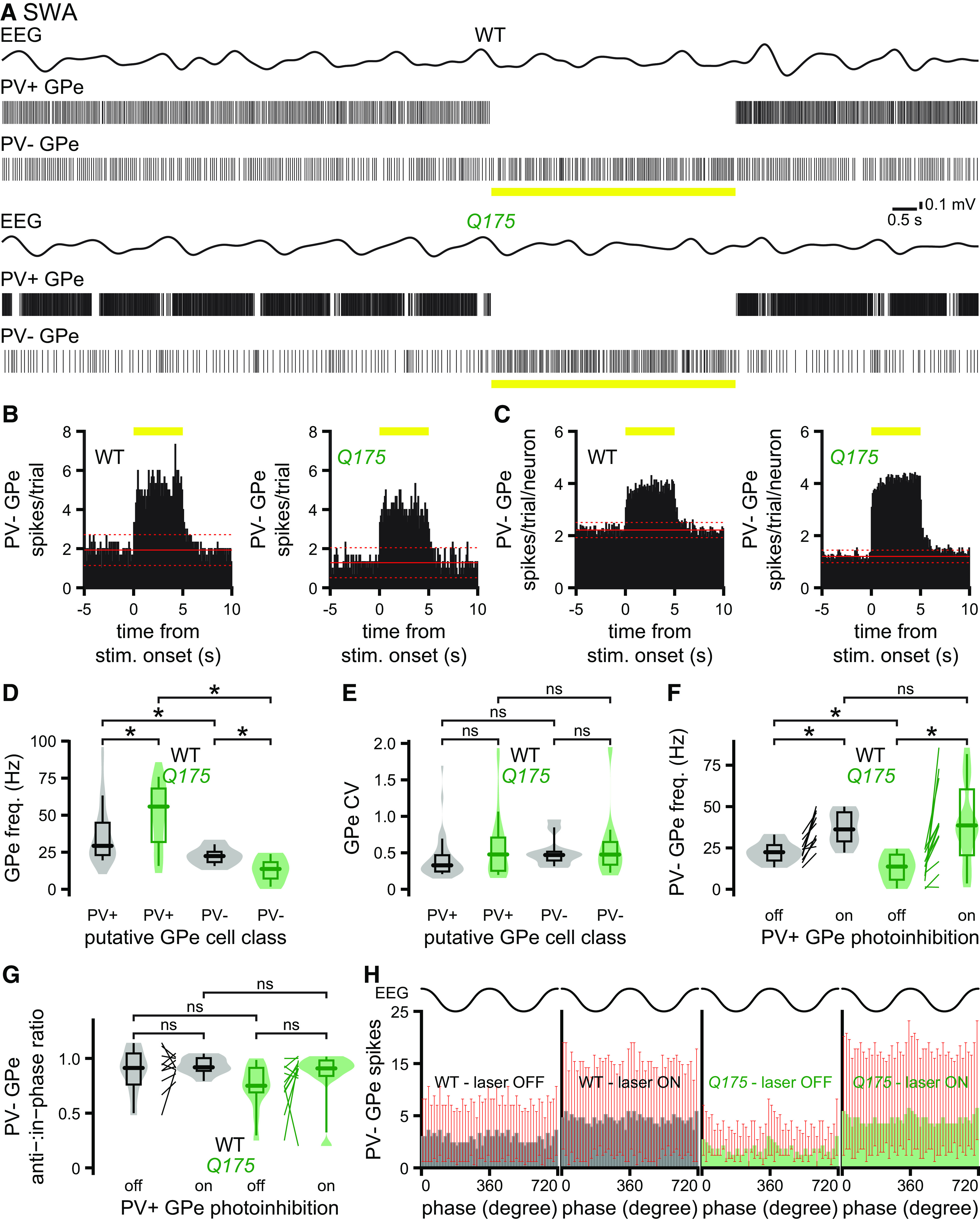
The hypoactivity of putative PV– GPe neurons in Q175 mice is alleviated by optogenetic inhibition of hyperactive PV+ GPe neurons. A, Representative examples of concurrent EEG (bandpass-filtered at 0.1-1.5 Hz) and GPe neuronal activity in Q175/WT X PV-cre mice during optogenetic inhibition (yellow bar) of PV+ GPe neurons. A–C, Optogenetic inhibition of PV+ GPe neuron activity rapidly and persistently disinhibited putative PV– GPe neurons in Q175 and WT mice. B, C, PSTHs of putative PV– GPe neuron activity in the absence and presence of PV+ GPe neuron optogenetic inhibition (yellow bar) (bin size, 100 ms; prestimulus mean, solid red line; ±2 SDs of prestimulus mean, dotted red line; B, PSTHs from representative neurons in A; C, population PSTHs). A–F, During cortical SWA, putative PV– GPe neurons were less active than PV+ GPe neurons in both WT and Q175 mice. Furthermore, the frequency of putative PV– GPe neuron activity in Q175 mice was lower than in WT mice (A, representative examples; D, population data). E, The precision of PV– GPe neuron activity was similar in WT and Q175 mice. Optogenetic inhibition of PV+ GPe neurons disinhibited putative PV– GPe neurons in WT and Q175 mice and eliminated the difference in firing frequencies between the two genotypes, arguing that GABAergic inhibition emanating from abnormally hyperactive PV+ GPe neurons is responsible for the relative hypoactivity of PV– GPe neurons in Q175 mice (A, examples; F, population data). G, H, Relative to cortical SWA, there were no differences in the phase of putative PV– GPe neuron activity in WT and Q175 mice, with or without optogenetic inhibition of PV+ GPe neurons. *p < 0.05.