. 2022 Mar 3;11:e74307. doi: 10.7554/eLife.74307

Figure 2. Corneal thickness is reduced in Npntkd corneas.

(A) Schematic of in ovo injection of viral constructs (green) to cover the anterior region of stage 8 chick embryo. Following 7 days of incubation, embryos were screened for GFP expression in the anterior eye region. Knockdown was verified by section in situ hybridization, which revealed reduced expression of Npnt in Npntkd cornea compared with control. (B–E) Hematoxylin and eosin staining showing control (B, D) and thinner Npntkd corneas (C, E). Statistical analysis on measurements taken from (N = 5 control and N = 4 Npntkd corneas) revealed (F) significant reduction in thickness of Npntkd corneas. **p<0.01. ep, corneal epithelium; st, stroma; en, corneal endothelium;. Scale bars: 100 μm (B, C), 100 μm (D, E).

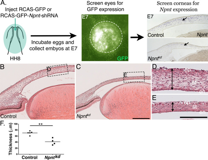
Figure 2.

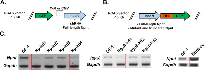
Figure 2—figure supplement 1. Diagram of replication-competent ASLV long terminal repeat with a splice acceptor (RCAS) vectors used for expression of either (A) GFP alone (control) or GFP together with inserts of shRNA or full-length nephronectin (Npnt).

Figure 2—figure supplement 1.

(B) The full-length or mutated and truncated versions of Npnt driven by the viral LTR promoter and GFP driven by the IRES promoter. (C) For RT-PCR, cells plated in 30 mm dishes were homogenized in 1 mL TRIzol, and RNA was isolated following the manufacturer’s protocol. RNA samples were treated with Turbo DNA-free kit (Invitrogen) to remove residual genomic DNA, and cDNA pools were generated using SuperScript First Strand System (Invitrogen). Semi-quantitative PCR was conducted using HotStart-IT Taq polymerase (Affymetrix). Knockdown efficiency was measured on 2% agarose gel using glyceralde-hyde-3-phosphate dehydrogenase (GAPDH) as a loading control. Red rectangles indicate the constructs that were chosen for the knockdown and overexpression experiments. Primers for RT-PCR are located in Supplementary file 1.