Figure 1.

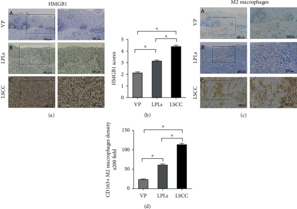
Immunohistochemical staining and semiquantitative evaluation of HMGB1 and CD163. (a) HMGB1 expression is scored as 2 in VP (a-A), 4 in LPLs (a-B), and 6 in LSCC (a-C) specimens. Original magnification, ×40 (left column), and selected areas (boxed areas), ×10. Semiquantitative evaluation of (b) HMGB1. To identify M2 macrophages in the tissue samples, an anti-CD163 antibody was used. (c) M2 macrophage density was greater in LSCCs (c-C) and LPLs (c-B) than in VP(c-A). Original magnification, ×40 (left column), and selected areas (boxed areas), ×10. Semiquantitative evaluation of (d) M2 macrophage density. Asterisks indicate statistically significant differences (∗P < 0.05).
