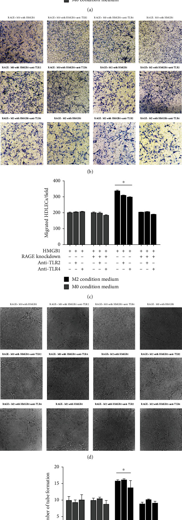
Figure 8.

HMGB1-TLR pathway does not significantly increase HDLEC proliferation, migration, and lymphangiogenesis on M2 macrophages. HDLEC incubated, respectively, with conditioned medium from RAGE+/- M0 macrophages preconditioned with HMGB1, RAGE+/- M0 macrophages preconditioned with HMGB1 and anti-TLR2/anti-TLR4 antibody, RAGE+/- M2 macrophages preconditioned with HMGB1, or RAGE+/- M2 macrophages preconditioned with HMGB1 and anti-TLR2/anti-TLR4 antibody. (a) CCK-8 assays were performed to assess the proliferation of HDLEC under various treatment conditions. HDLEC were seeded into upper chamber of Transwell plates and counted by light microscopy after 6 h. Representative (b) micrographs and (c) cell counts for migration are shown. The formation of lymphatic vessels was counted in Matrigel after 6 h. Representative (d) micrographs and (e) formation of lymphatic vessels counts are shown. Data are presented as the means ± SD and are representative of 3 independent experiments (∗P < 0.05).
