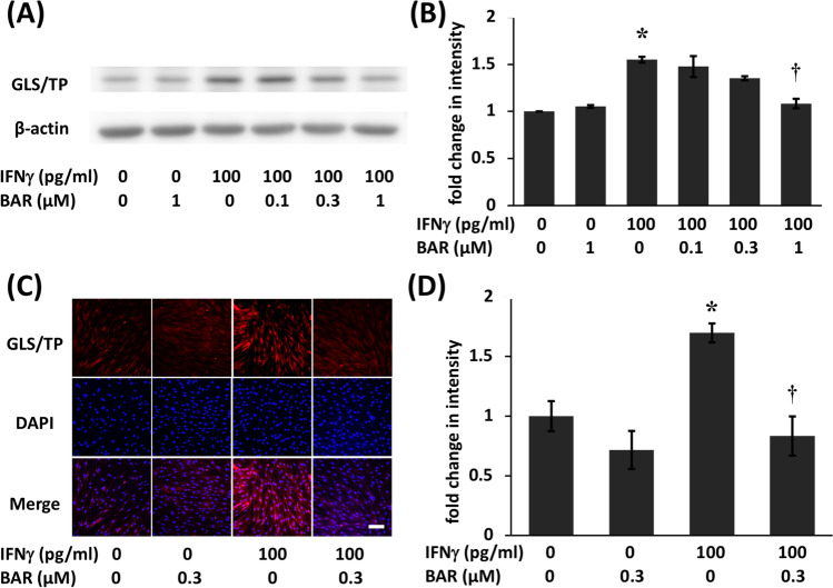
Fig. 2.
Baricitinib suppressed interferon (IFN)γ-induced gliostatin/thymidine phosphorylase (GLS/TP) protein expression in fibroblast-like synoviocytes (FLSs) from RA patients (RA). A Confluent FLSs were incubated in the presence or absence of 0.1 to 1 μM baricitinib for 6 h and then with 100 pg/ml IFNγ for 18 h. GLS/TP protein levels were assessed by Western blotting. B Band density was measured using ImageJ. The statistical significance of differences between groups was calculated by Tukey’s multiple comparisons test. Compared with controls: *P < 0.001. Compared with samples treated without baricitinib: †P < 0.001. Immunocytochemical detection of GLS/TP in FLSs from RA patients. C FLSs were treated with or without 0.3 μM baricitinib for 6 h before being incubated with or without IFNγ (100 pg/ml) for 18 h, after which they were immunostained with a GLS/TP antibody (red). The cell nuclei were stained with 4′,6-diamidino-2-phenylindole (DAPI) (blue). The scale bar represents 100 μm. Control FLSs were cultured without additional agents. D The total intensity of immunostaining in a random field was quantified by ImageJ, and the number of cells in the field was counted. The data (intensity/cell) are presented as the mean ± standard error of the mean (SEM) of five determinations. The statistical significance of differences between groups was calculated by one-way analysis of variance (ANOVA) and Tukey’s multiple comparisons test. Compared with controls: *P < 0.05. Compared with samples treated with IFNγ alone: †P < 0.05. BAR, baricitinib

