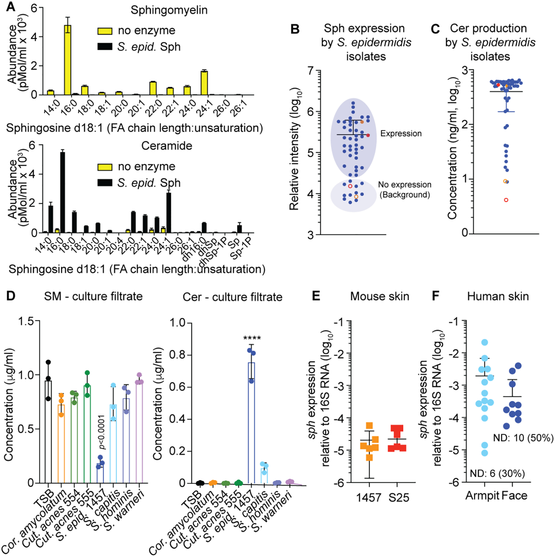
Fig. 2. Enzymatic activity, species-wide and in-vivo expression of Sph.

A, Cleavage of sphingomyelin from human keratinocytes by purified Sph (100 μg/ml). Shown is the Tandem-MS analysis of the major sphingomyelin type with d18:1 sphingosine, producing type-2 ceramide. FA, fatty acid; Sp, sphingosine (d18:1) of undetermined FA chain length; dh, dihydro (d18:0 instead of d18:1); P, phosphate. n=3. See Fig. S2 for results with other tissue sources using RP-HPLC/ESI-MS. B, In-vitro expression of Sph by Western blot and relative densitometric analysis in a collection of S. epidermidis isolates. Filled colored circles show data obtained with strains 1457 (red) and S25 (orange), open circles those with strains 1457 Δsph (red) and S25 Δsph (orange). See Fig. S4 for Western blot images. C, Production of ceramides by culture filtrates of a collection of S. epidermidis isolates. Filled colored circles show data obtained with strains 1457 (red) and S25 (orange), open circles those with strains 1457 Δsph (red) and S25 Δsph (orange). D, SM degradation and Cer production by other skin-colonizing bacteria compared to S. epidermidis. n=3. Statistical analysis is by 1-way ANOVA with Dunnett’s post-test via data obtained with TSB. E, Expression of the sph gene on mouse skin associated with the S. epidermidis strains used in this study. Expression was determined 1 day after association. n=6/group. F, Expression of the sph gene in swabs obtained from armpits and faces of human volunteers. n=20. Computation of mean and SD includes 0 values from individuals without detectable expression (ND). A-F, Error bars show the mean ± SD. S. epid., S. epidermidis; Cut. acnes, Cutibacterium acnes; Cor. amycolatum, Corynebacterium amycolatum.
