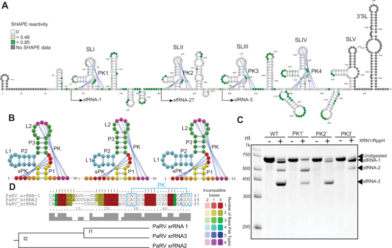
Fig. 3. cISFs PaRV contains multiple copies of divergent class 1b xrRNAs.
A Secondary structure of PaRV 3’UTR generated using SHAPE-assisted folding. Colour intensity indicates NMIA reactivity. Values are the means from 3 independent experiments, each with technical triplicates. SL – stem loop, PK -pseudoknot. B Secondary structure of PaRV xrRNAs. Each structural element is shown in colour. C In vitro XRN1 resistance assay with WT and mutated PaRV 3’UTRs. PK1’, PK2’ and PK3’ mutations were introduced into the terminal loop regions of SLI, SLII and SLIII, respectively, and represented the CAC - > GUG change in the PK-forming regions. Image is representative from three independent experiments that showed similar results. D Structure-based alignment of PaRV xrRNAs was performed using LocARNA.

