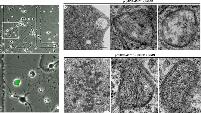
Figure 6.
Correlative light electron microscopy shows improvement of mitochondrial ultrastructure upon NMN treatment. (a) a representative image of motor cortex neurons cultured on a gridded glass bottom 35 mm dish; boxed area enlarged to highlight presence of GFP expressing CSMN; (b) representative electron micrograph of an individual CSMN from prpTDP-43A315T-UeGFP mouse showing mitochondrial ultrastructural defects, images of mitochondrion are enlarged to right; and (c) representative electron micrograph of an individual CSMN from prpTDP-43A315T-UeGFP treated with 1 µM NMN showing improved mitochondrial ultrastructure, images of mitochondrion are enlarged to right. Scale bar = 1 µm.

