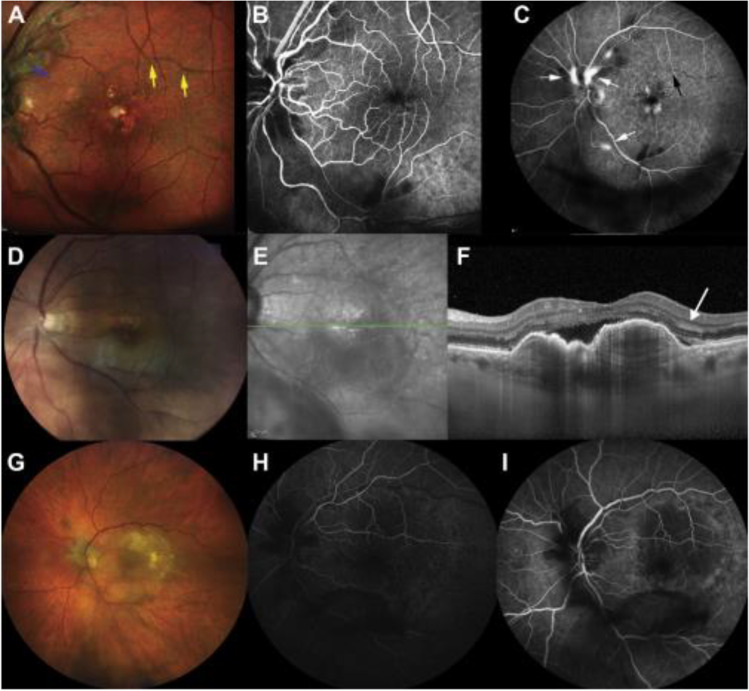
Fig. 4.
Three representative cases demonstrating the spectrum of ocular findings related to IOI and occlusive retinal vasculitis. Case 1 (A–C): An 88-year old woman was diagnosed with retinal vasculitis in her left eye at 6 weeks after bilateral intravitreal brolucizumab injection. Color fundus photograph (A) reveals multiple intra-arterial foci of gray material (yellow arrow) and retinal whitening extending from the optic nerve along the superotemporal arcade (blue arrow). Fluorescein angiography (B early, C late) shows delayed flow along the inferotemporal arcade, with late focal staining of the retinal arteries (white arrow). A region of nonperfusion is noted superior to the fovea (black arrow) corresponding to the foci of intra-arterial gray material in 2A. Case 2 (D–F): An 80-year-old woman presented with reduced vision and a superior scotoma at 7 days after her second brolucizumab injection. Fundus photograph (D) shows retinal whitening involving the inferior macula, arterial sheathing, and focal interruptions of the blood column within an inferotemporal macular branch retinal artery. Near-infrared (E) and OCT (F) show subretinal fluid that was improved from prior OCT evaluations and intraretinal foci of hyperreflectivity (white arrow). Case 3 (G–I): A 75-year-old woman had persistent subretinal fluid despite 18 previous anti-VEGF injections (14 aflibercept/4 ranibizumab), comprising the reason to switch to brolucizumab. She presented with floaters and reduced vision and was diagnosed with IOI and occlusive retinal vasculitis 30 days after her first brolucizumab injection. Fundus photograph (G) shows multiple cotton wool spots around the optic nerve and perimacular and subtle periarterial whitening. There is some vitreous opacity along the inferotemporal arcade. Fluorescein angiography (H, early 28 s) shows globally delayed retinal arterial filling, notable around the optic nerve. At 68 s (I), there remains delayed arterial filling around the optic nerve and inferiorly, and blockage from vitreous opacity. A to C courtesy of Haug et al. and D to G courtesy of Baumal et al. Reprinted from Ophthalmology Retina, 5(6), Baumal CR, Bodaghi B, Singer M et al. Expert Opinion on the Management of Intraocular Inflammation, Retinal Vasculitis, and Vascular Occlusion after Brolucizumab Treatment, 519–527, Copyright (2021), with permission from Elsevier

