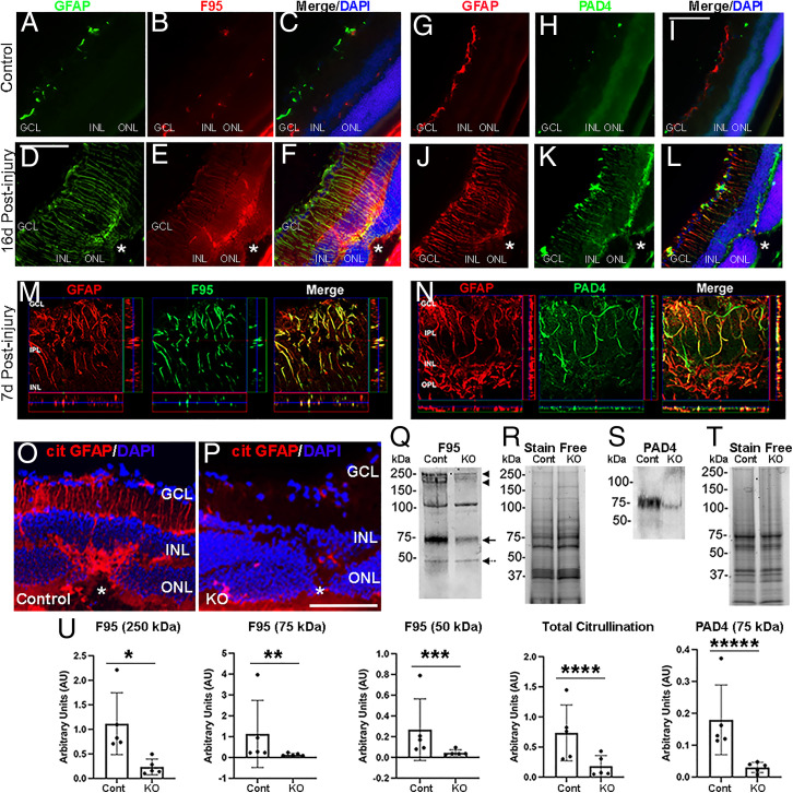
Fig. 1.
Citrullination and PAD4 in reactive MG after laser injury. (A–F) Representative images of retinal sections from uninjured and postinjured mice immunostained for GFAP (green) and citrullination (F95, red). Nuclei were visualized with DAPI (blue). Asterisks demark laser injury site. (M) Orthogonal projections of confocal z-stacks stained for GFAP (red) and F95 (green) at high magnification. (G–L) Representative images of retinal sections from uninjured and postinjured mice immunostained for GFAP (red) and PAD4 (green). (N) Orthogonal projections of confocal z-stacks stained for GFAP (red) and PAD4 (green). (Scale bars, 105 μm.) (O and P) Representative images of retinas from laser injured control and PAD4cKO (KO) mice immunostained for cit-GFAP. GCL, ganglion cell layer; INL, inner nuclear layer; ONL, outer nuclear layer. WB analysis of retinal extracts subjected to denaturing and reducing conditions from laser-injured control and KO retinas probed for citrullination (F95; Q) and PAD4 (S). (U) Quantitation of F95 reactive bands and PAD4 were normalized to stain-free gels (R and T), respectively (n = 5 blots; error bars are SD of mean; *P = 0.004, **P = 0.0159, ***P = 0.0159, ****P = 0.0278, *****P = 0.004) using one-way ANOVA.

