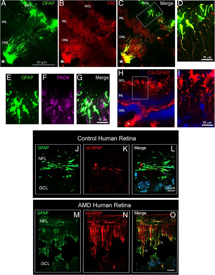
Fig. 2.
Citrullinated GFAP in MG endfeet of JR5558 mouse and human wet-AMD diseased retinase. Localization of cit-GFAP in JR5558 mouse retinas and human wet-AMD macula. One-month-old JR5558 mouse retinas were immunostained for GFAP, citrullination (F95), cit-GFAP, and PAD4. Lesion-specific staining for GFAP (A; green) overlaps with staining with F95 (B; red) and is seen in the merged image (C). Representative higher magnification images show F95 colocalization with GFAP in endfeet (D). Representative higher magnification images of GFAP staining (E; green) and PAD4 (F; magenta) showing colocalization (G). Cit-GFAP staining in a 2-mo-old JR5558 mouse retina (H) with higher magnification revealed at endfeet (I). Asterisks mark the site of spontaneous retinal lesions. (J–O) Representative tissue sections from 89-y-old wet-AMD and age-matched control donor maculae stained for GFAP (green) and cit-GFAP (red). The endfeet expression of cit-GFAP in wet-AMD macula is revealed in high magnification images showing overlap with GFAP staining (M–O; arrowheads), whereas staining in controls is low (K) and overlaps minimally with GFAP (L). NFL, nerve fiber layer; GCL, ganglion cell layer; INL, inner nuclear layer; ONL, outer nuclear layer. (Scale bar, 10 μm.).

