Fig. 3.
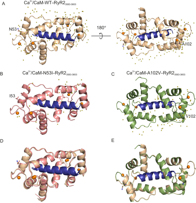
The arrhythmogenic N53I mutant causes subtle changes in the three-dimensional structure of Ca2+/CaM–RyR23583-3603. (A-C) Cartoon representation of the crystal structures of Ca2+/CaM proteins in complex with RyR2 peptide. (A) Ca2+/CaM-WT–RyR23583-3603 (PDB 6XXF). (B) Ca2+/CaM-N53I–RyR23583-3603 (PDB 6XY3). (C) Ca2+/CaM-A102V–RyR23583-3603 (PDB 6XXX). (D,E) Alignments of Ca2+/CaM-WT–RyR23583-3603 with Ca2+/CaM-N53I–RyR23583-3603 (D) or Ca2+/CaM-A102V–RyR23583-3603 (E) complex structures. Ca2+ ions are shown as orange spheres and water molecules as olive spheres. The WT and mutant residues are shown in stick representation. CaM-WT is displayed in beige, CaM-N53I in salmon, CaM-A102V in green and RyR23583-3603 peptide in blue. Images were generated using PyMOL software.

