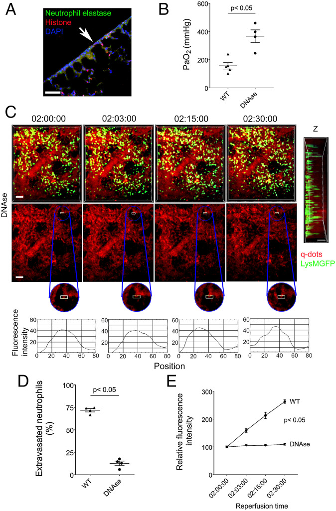
Fig. 7.
DNase treatment prevents disruption of subpleural capillary network. (A) Colocalization of neutrophil elastase, histones, and DAPI in DNase-treated wild-type (WIS) grafts by immunofluorescent staining. The arrow points to pleural surface (Scale bars: 100 μm). (B) Arterial blood oxygenation 2 h after transplantation of B6 wildtype (WIS) into syngeneic recipients, without and with DNase treatment. (C) Time lapse intravital two-photon imaging of neutrophils (green) (Top [Scale bars: 30 μm]), quantum dots (red) that were injected intravenously (Middle [Scale bars: 30 μm]), quantification of disruption of vascular integrity as evidenced by extravascular quantum dot signal (boxed region and kymographs), and side projections of z stacks (Scale bar: 20 μm) of wild-type lungs (WIS) after transplantation into DNase-treated syngeneic recipients. (D) Percentage of extravasated neutrophils and (E) comparison of extravascular quantum dot intensity in subpleural space of B6 wild-type lungs (WIS) over time after transplantation into syngeneic recipients that received no treatment or were treated with DNase. The data in (B), (D), and (E) represent the mean ± SEM (n = 4). The Left side of z stack in (C) denotes pleural surface. Statistical analysis for (E) is for last time point.

