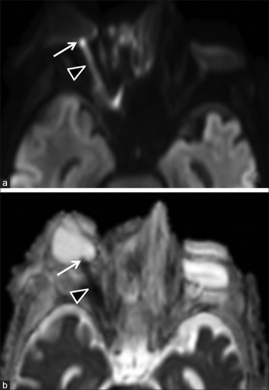
Figure 1.

(a) Diffusion-weighted image (DWI) and (b) corresponding apparent diffusion coefficient (ADC) map show restricted diffusion in the intraocular (arrow) and intraorbital (arrowhead) segments of the right optic nerve, representing anterior ischemic optic neuropathy (AION) and posterior ischemic optic neuropathy (PION), respectively. Left optic nerve appears normal
