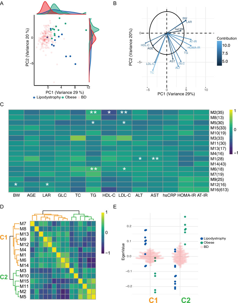
Fig. 1.
Metabolic signatures in the obese and lipodystrophy groups. a Principal component analysis (PCA) of three groups: obese, green; lipodystrophy, blue; and blood donors (BD), light red. PCA was performed using the parameters below. b Representation of PCA loadings on: age, weight (BW), body mass index (BMI), leptin-adiponectin ratio (LAR), glucose (GLC), triglycerides (TG), total cholesterol (TC), high-density lipoprotein (HDL-C), low-density lipoprotein (LDL-C), alanine amino-transferase (ALT), aspartate amino-transferase (AST), Homeostatic Model Assessment for Insulin Resistance (HOMA-IR) and adipose tissue insulin resistance (AT-IR) indexes and high-sensitivity C-reactive Protein (hsCRP). Colour and arrow length scale represent contribution to variance on the first two principal components. c Metabolite module-trait associations using WGCNA consensus analysis and 988 metabolites. Each row corresponds to a module eigen metabolites (ME) and each column to a parameter. Number of metabolites in each module is indicated in brackets. Cell colour represents Pearson’s correlation as shown by legend. Significance is annotated as follows: *p ≤ 0.05; **p ≤ 0.01; ***p ≤ 0.001; ****p ≤ 0.0001 (Fisher’s test p value corrected for multi-testing). d Heatmap of extreme phenotype groups’ MEs adjacencies in the consensus MEs network. The heatmap is colour-coded by adjacency, yellow indicating high adjacency (positive correlation) and blue low adjacency (negative correlation). e Beeswarm plot using average MEs per cluster presented in (d)

