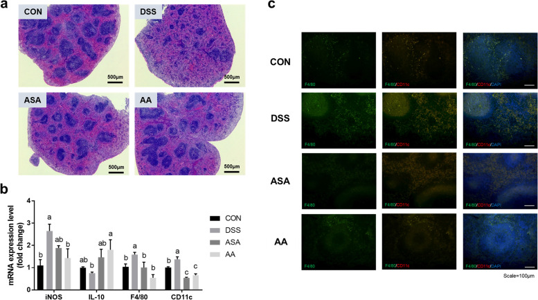
Fig. 6.
A. argyi treatment enhanced immune responses in the splenic tissue of mice with DSS-induced colitis. a The histological image of spleen tissue. Mouse spleen tissue sections were stained with H&E (scale bar = 500 μm). b mRNA expression of inflammation-related markers and M1 macrophage markers (iNOS, IL-10, F4/80, and CD11c). The mRNA level was normalized to that of GAPDH. c Immunohistochemistry against F4/80 and CD11c was performed in mouse splenic tissue (scale bar = 100 μm). Data are presented as mean ± SEM from three independent experiments. Different superscripts indicate significant differences at P < 0.05. CON, the mice group was treated with pure water; DSS, the mice group was treated with 3% DSS solution; ASA, the mice group was treated with 3% DSS solution and 100 mg kg-1 day-1 of 5-amino salicylic acid (5-ASA); AA, the mice group was treated with 3% DSS solution and 200 mg kg-1 day-1 of A. argyi extract

