Fig. 1.

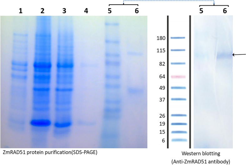
SDS-PAGE gel and western blot showing purification of RAD51A1. An SDS-PAGE gel is shown on the left, and a western blot probed with an α-ZmRAD51 antibody from rabbit is shown on the right. Gel lanes represent (1) supernatant, (2) pellet, (3) flow through, (4) wash, (5) marker, and (6) eluted protein. In the western blot on the right, lane (6) contains the eluted RAD51 protein probed with an α-ZmRAD51 antibody from rabbit. The RAD51A1 dimer band (92 KDa), indicated by an arrow, can be seen more clearly than the monomer band (46 KDa)
