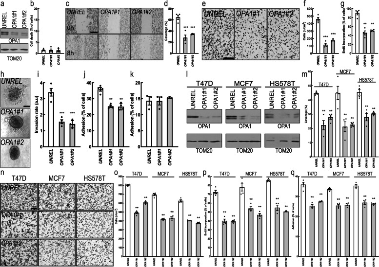
Fig. 2.
OPA1 is required for breast cancer cells hallmarks in vitro. a MDA-MB-231 cells were transfected with the indicated siRNA and lysed after 72 h. Equal amounts of proteins were separated by SDS-PAGE and immunoblotted with the indicated antibodies. b Quantification of apoptosis rate in MDA-MB-231 transfected with the indicated siRNA for 72 h determined by annexin V/propidium iodide label by flow cytometry. n = 4 independent experiments. c MDA-MB-231 cells were transfected with the indicated siRNA and after 72 h a scratch-wound assay was performed. Representative brightfield images were acquired at the indicated time points. Scale bar: 250 μm. d Quantification of cell migration after 6 h in n = 4 independent experiments as in (b). ***: p < 0.0001. e Representative brightfield images of MDA-MB-231 cells transfected with the indicated siRNA in a Boyden chamber. Scale bar: 250 μm. f Quantification of cell migration in a Boyden chamber after 6 h in experiments as in (d). n = 4 independent experiments. ***: p < 0.0001. g Quantification of MDA-MB-231 proliferation upon transfection with the indicated siRNA determined by BrdU incorporation. n = 4 independent experiments. **: p < 0.001. h Representative brightfield images of MDA-MB-231 spheroïds transfected with the indicated siRNA. Scale bar: 250 μm. i Quantification of cell invasion after 6 h in experiments as in (h). n = 4 independent experiments. ***: p < 0.0001. j Quantification of cell adhesion on fibronectin for 1 h of MDA-MB-231 transfected with the indicated siRNA for 72 h. n = 4 independent experiments. ***: p < 0.001. k Quantification of cell adhesion on gelatin for 1 h of MDA-MB-231 cells transfected with the indicated siRNA for 72 h. n = 3 independent experiments. l Equal amounts of protein from breast cancer T47D, MCF7 and HS578T cells transfected for 72 h with the indicated siRNA were separated by SDS-PAGE and immunoblotted with the indicated antibodies. m Quantification of migration after 6 h in a scratch wound assay of T47D, MCF7 and HS578T cells transfected with the indicated siRNA for 72 h. n = 4 independent experiments. **: p < 0.001. n Representative brightfield images of T47D, MCF7 and HS578T cells transfected for 72 h with the indicated siRNA in a Boyden chamber 6 h after induction of migration. Scale bar: 250 μm. o Quantification of cell migration in experiments as in (n). n = 4 independent experiments. **: p < 0.001. p Quantification of proliferation of T47D, MCF7 and HS578T transfected with the indicated siRNA determined by BrdU incorporation. n = 4 independent experiments. **: p < 0.001. q Quantification of cell adhesion on fibronectin for 1 h of T47D, MCF7 and HS578T cells transfected with the indicated siRNA for 72 h. n = 4 independent experiments. **: p < 0.001

