
Fig. 5. TSLP stimulates T cells to promote sebum production and increases T cells in the skin.
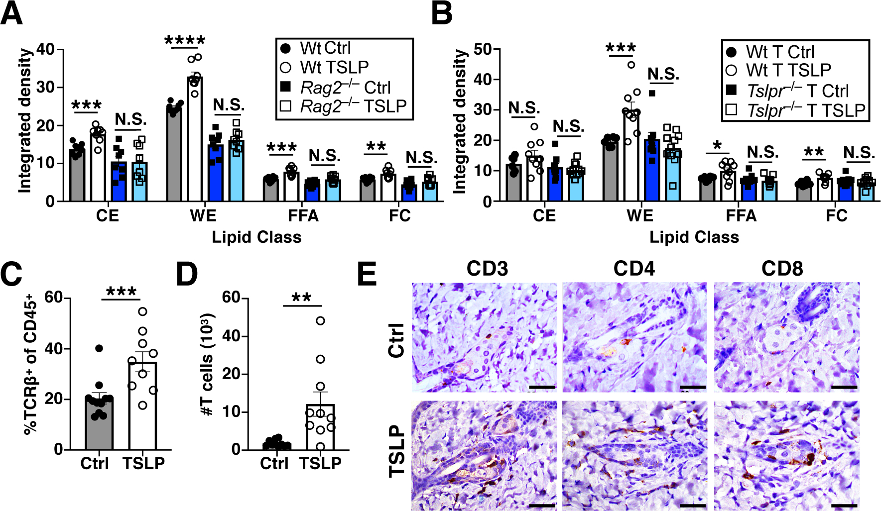
(A) TLC quantification of hair lipids from Wt and Rag2−/− mice, 2 weeks post AAV (n=8 mice/group, pooled from 3 independent experiments). t-test. (B) TLC quantification of hair lipids from Rag2−/− mice reconstituted with Wt or Tslpr−/− T cells, 2 weeks post AAV (n=6–9 mice/Wt T cell group, pooled from 2 independent experiments, and n=9–11 mice/Tslpr−/− T cell group, pooled from 3 independent experiments). t-test. (C and D) Percentage and number of skin T cells by flow cytometry, 10 days post AAV (n=9–11 mice/group, pooled from 3 independent experiments). t-test. (E) Skin CD3, CD4, and CD8 immunohistochemical staining, 10 days post AAV. Scale bars=40 μm. N.S., P≥0.05, *P<0.05, **P<0.01, ***P<0.001, ****P<0.0001. Data are mean ± SEM.