FIG. 1.
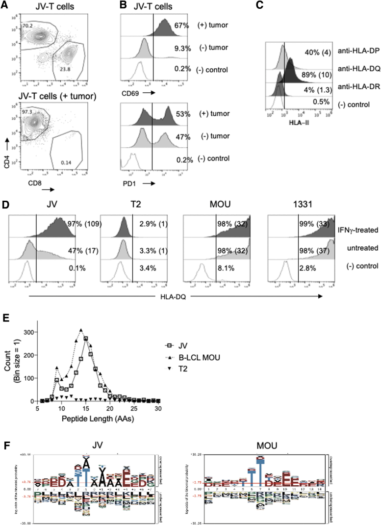
HLA-DQ-eluted peptides in JV ATC cells. (A, B) Characterization of peripheral T cells isolated from JV patient as determined by flow cytometry. (A) Frequencies of CD4:CD8 in peripheral JV-T cells (top) or in in vivo expanded JV-T cells after exposure to autologous tumor cells in vitro (bottom). (B) Expression of CD69 (top) or PD1 (bottom) in JV-T cells before or after exposure to autologous tumor cells. (C) Surface expression of MHC II molecules in JV tumor cells was determined by flow cytometry. The gates for positively stained cells were determined by staining with a secondary antibody only and were marked with vertical lines within the plots. The numbers in the parenthesis are the fold increases in mean fluorescence intensity of each MHC II expression. (D) The expression levels of HLA-DQ in uninduced and IFNγ-induced conditions (2e4 IU/mL for 48 hours) were determined in JV, T2, B-LCL MOU, and 1331 cells using SPV-L3 antibody. HLA-DQ histograms analyzed with the 1a3 antibody presented a similar profile to SPV-L3 (Supplementary Fig. S2). JV cell HLA-DQ haplotypes were HLA-DQA1*0201, HLA-DQB1*0202/HLA-DQB1*0302. MOU and 1331 B-LCL lines had DQ2.2 (HLA-DQA1*0201/HLA-DQB1*0202) and DQ8.1 (HLA-DQA1*0301/HLA-DQB1*0302), respectively. T2 cells were deficient in MHC II expression including HLA-DQ. (E) Peptide length distribution of HLA-DQ-eluted peptides from MOU, T2, and IFNγ-induced JV cells was shown as a histogram. (F) Sequence logos of eluted peptides isolated from JV-ATC (15 a.a., left) and MOU (14 a.a., right) were generated using pLogo generator (38). ATC, anaplastic thyroid cancer; B-LCL, B lymphoblastic cell line; HLA, human leukocyte antigen; MHC, major histocompatibility complex.

