Figure 7.
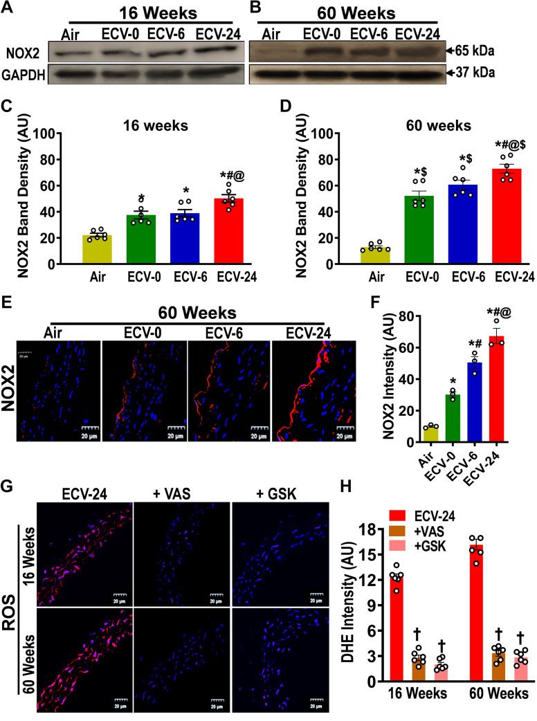
Expression of NADPH oxidase 2 in the aorta. NADPH oxidase 2 (NOX2) levels were measured by Western blotting (A–D) and immunofluorescence (E and F) from mice exposed to 16 and 60 wk of either air or electronic cigarette (e-cig) vape generated from liquid containing nicotine (NIC) 0 mg/mL (ECV-0), 6 mg/mL (ECV-6), or 24 mg/mL (ECV-24). A and B: immunoblots of NOX2. C and D: quantitation of NOX2 band density in A and B, respectively, showing exposure time- and NIC-dependent increases in NOX2 expression. E: aortic sections were incubated with primary antibody against NOX2 followed by corresponding secondary fluorescent tagged antibody (red), with DAPI nuclear stain (blue). F: quantitation of red fluorescence in E, showing a similar response as in D. G: aortic sections from ECV-24-exposed mice were incubated with the superoxide probe dihydroethidium (DHE; red) and DAPI (blue), with or without NADPH oxidase inhibitors 3-benzyl-7-(2-benzoxazolyl)thio-1,2,3-triazolo[4,5-d]pyrimidine (+VAS) or GSK2795039 (+GSK), and visualized by confocal microscopy. H: quantitation of the superoxide-derived red fluorescence in G, showing that NOX2 inhibitors decreased superoxide in ECV-24-exposed aortic sections. Thus, induction of NOX2 in the endothelium is a major source of the ECV-stimulated superoxide seen in all ECV-exposed groups, and its induction increased with exposure duration and NIC content. For C and D, data are presented as means ± SE of 6 mice, whereas for F and H, n = 3 or 6 mice, respectively. Analysis was done using two-way ANOVA followed by Bonferroni multiple-comparisons test. The differences were considered statistically significant at P ≤ 0.05. *Significant from air-exposed controls at P < 0.05; #significant from ECV-0 at P < 0.05; @significant from ECV-6 at P < 0.05; $significant from the same exposure at 16 wk at P < 0.05. †significant different from ECV-24-exposed sections stained with DHE.

