Figure 3.
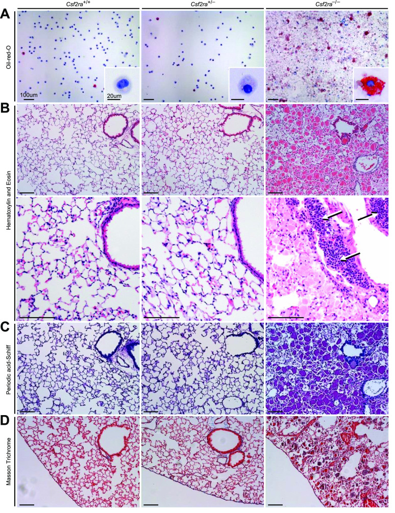
Effects of Csf2ra gene disruption on bronchoalveolar lavage (BAL) cytology and lung histology. A: examination of BAL after Oil-red-O staining revealed normal cytology in Csf2ra+/+ and Csf2ra+/– mice and abnormal cytology, numerous large, red macrophages (indicating cholesterol ester-rich inclusions) and “dirty”-appearing extracellular debris in Csf2ra–/– mice. Scale bar is 100 µm in main panel and 20 µm in inset. B: lung histology was normal in Csf2ra+/+ and Csf2ra+/– mice and abnormal in Csf2ra–/– mice; hematoxylin and eosin staining revealed well-preserved alveoli filled with homogeneous eosinophilic sediment (second row, right) and lymphocytosis (arrows) in perivascular distribution (third row, right). Scale bar is 100 µm. C: periodic acid-Schiff staining was normal in Csf2ra+/+ and Csf2ra+/– mice and abnormal histology in Csf2ra–/– showing a pattern typical of hereditary pulmonary alveolar proteinosis (hPAP) (fourth row, right). Scale bar is 100 µm. D: Masson trichrome staining did not reveal any significant fibrosis in either Csf2ra+/+, Csf2ra+/–, or Csf2ra–/– mice (bottom row). Scale bar is 100 µm.

