
Figure 1.
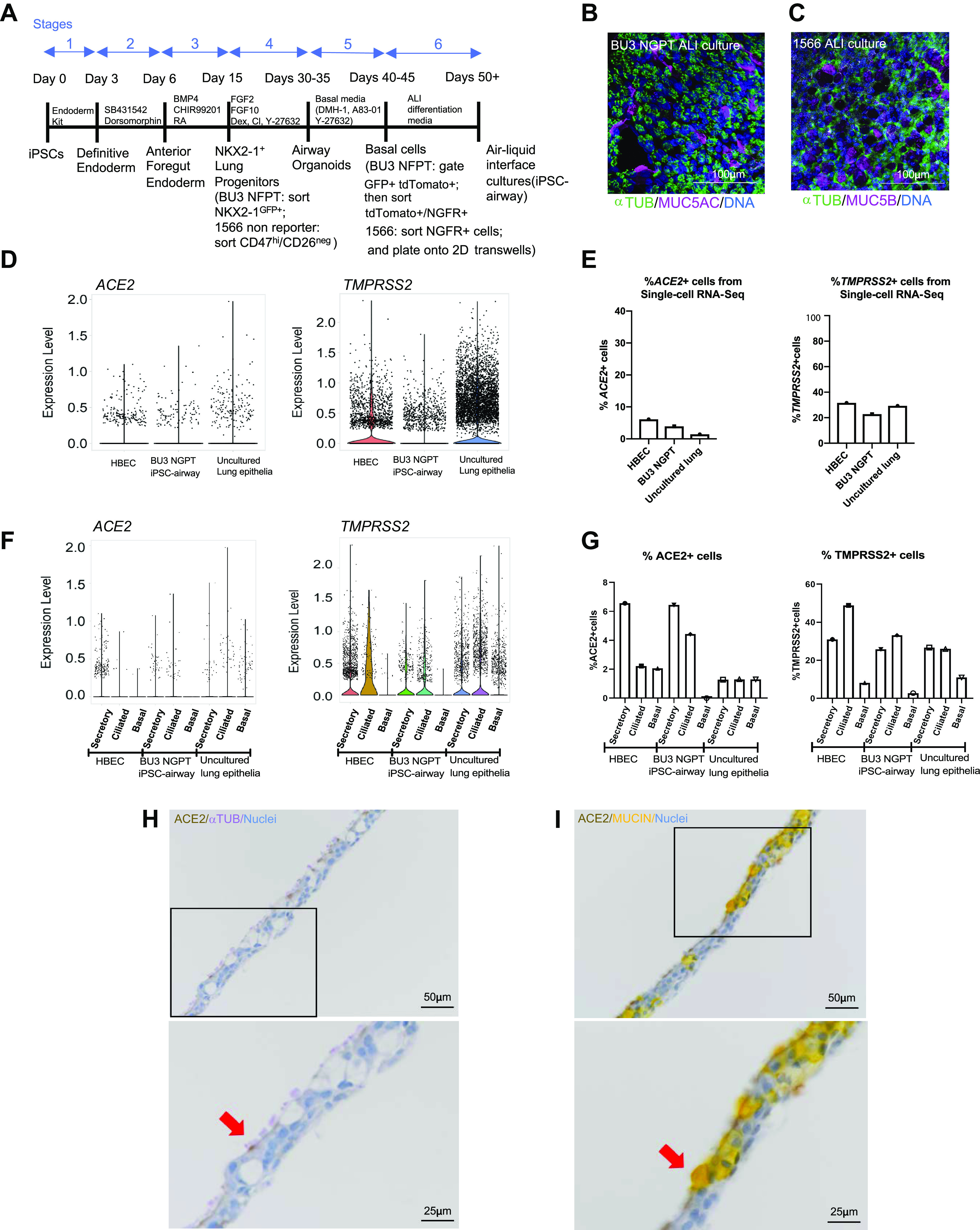
iPSC-derived airway cells express SARS-CoV-2 entry factors angiotensin converting enzyme 2 (ACE2), and transmembrane serine protease 2 (TMPRSS2). A: schematic of the six-stage differentiation protocol of generating iPSC airway. B: immunofluorescence analysis of BU3 NGPT iPSC airway stained with anti-α-TUBULIN and mucin 5AC (MUC5AC) (scale bar = 100 µm). Nuclei are stained with HOECHST (blue). C: immunofluorescence analysis of 1566 iPSC airway, stained with anti-α-TUBULIN and mucin 5B (MUC5B) (scale bar = 100 µm). Nuclei are stained with DAPI (blue). D–G: scRNA-seq analysis of human bronchial epithelial cells (HBEC)40, iPSC airway (BU3 NGPT) (40), and freshly isolated uncultured lung epithelia (42). D: violin plots of ACE2 and TMPRSS2 expression. E: percentage of ACE2- and TMPRSS2-positive cells in each data set (40). F: violin plots of ACE2 and TMPRSS2 expression by cellular type in each data set. G: comparison of the percentage of ACE2- and TMPRSS2-positive secretory, multiciliated, and basal cells in each data set. H and I: immunohistochemistry staining showing the localization of ACE2 (DAB), α-TUBULIN (purple, left) and MUCIN (yellow, right) in iPSC-derived airway (BU3 NGPT) counterstained with hematoxylin (×20, scale bar = 50 µm). Bottom: zoomed-in images of the black box in the upper panels. The red arrows indicated cells coexpressing ACE2/α-TUBULIN (left) and ACE2/MUCIN (right; scale bar = 25 µm). ACE2, human angiotensin-converting enzyme; ALI, air-liquid interface; HBECs, human primary bronchial epithelial cells; iPSC, induced pluripotent stem cell; TMPRSS2, transmembrane protease, serine 2.