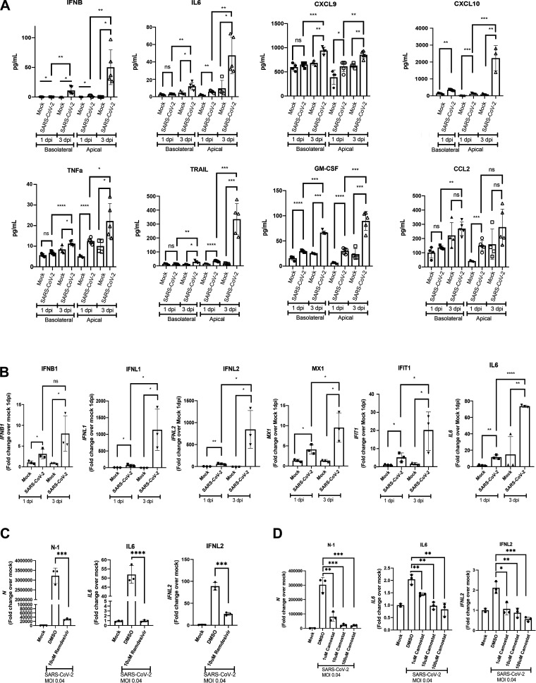
Figure 4.
iPSC airway infected with SARS-CoV-2 secrete inflammatory cytokines and chemokines and detects antiviral drug effects. A: luminex analysis of apical washes and basolateral media collected from iPSC airway (BU3 NGPT, cultures; n = 3 Transwells). B: RT-qPCR of select genes iPSC airway (BU3 NGPT) infected with SARS-CoV-2 (MOI 4) and harvested at 1 and 3 dpi with their respective mock-infected samples (n = 3 Transwells). Fold-change expression compared with mock (2−ddCt) after 18S normalization is shown. C and D: RT-qPCR of N, IL6, IFNL2 gene expression of mock-infected and SARS-CoV-2-infected (MOI 0.04) iPSC airway (BU3 NGPT) at 2 dpi pretreated with vehicle control (DMSO) or remdesivir (10 μM; C) or vehicle control (DMSO) or camostat mesylate (TMPRSS2 inhibitor, 1, 10, 100 μM; D), as indicated (n = 3 Transwells for both C and D). Statistical significance was determined as P value of <0.05 using Student’s t test (*P ≤ 0.05; **P ≤ 0.01; ***P ≤ 0.001; ****P ≤ 0.0001). iPSC, induced pluripotent stem cell; MOI, multiplicity of infection.

