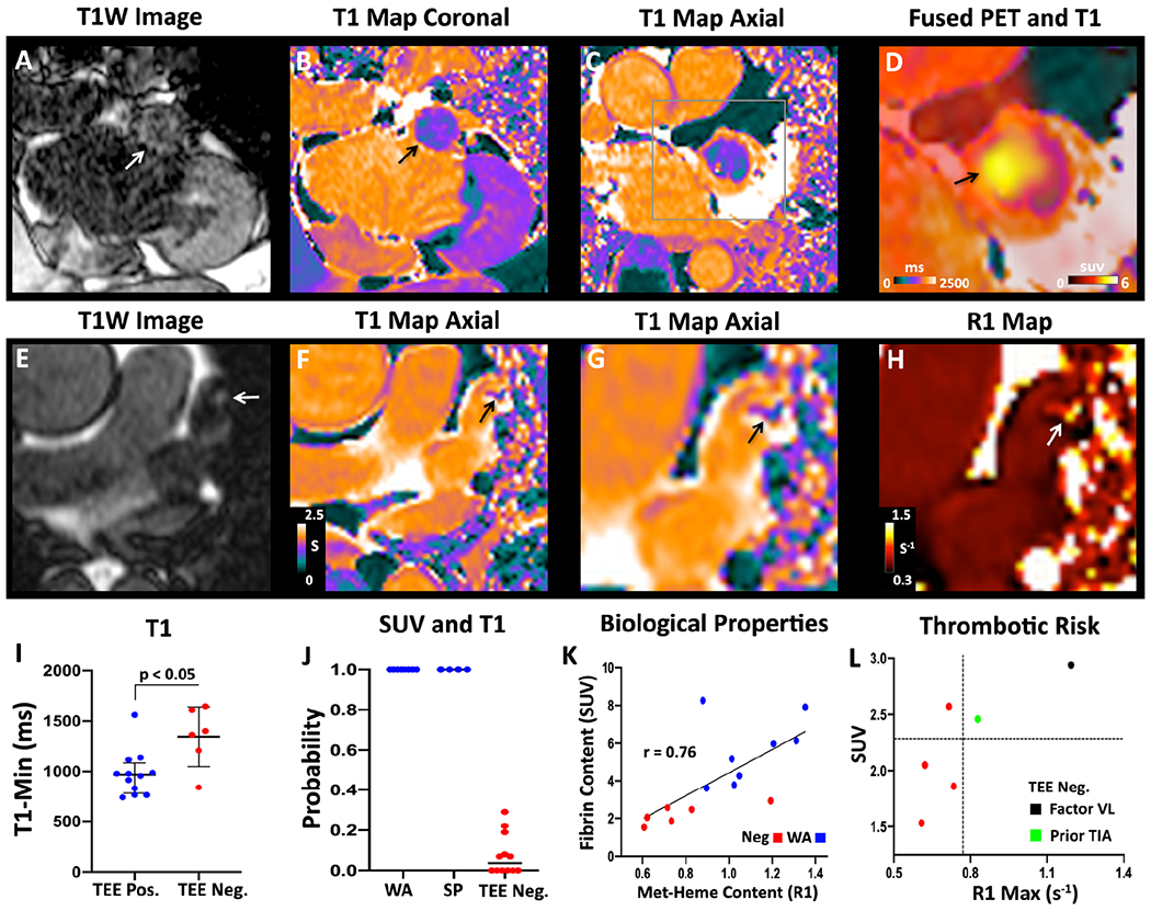
Figure 3. Integrated analysis of magnetic relaxation (T1) and SUVMAX in the LAA.

(A-D) Subject with a LAA closure device. (A) T1-weighted image in the coronal plane showing a hyperintense region in the LAA (white arrow). (B) T1 map in the same view demonstrating low values (green-purple; black arrow) in the LAA, similar to the T1 of myocardium (purple) but far lower than the blood pool (orange). (C) Axial T1 map confirms the presence of short-T1 (paramagnetic) species in the LAA, consistent with methemoglobin (met-heme) accumulation in thrombus. (D) Magnified view of the axial T1map fused with the [64Cu]FBP8 PET image demonstrates a high degree of the overlap between low T1 and high [64Cu]FBP8 uptake. (E-H) Spontaneous LAA thrombus. (E) Axial T1W image showing a hypertintense focus (arrow) in the LAA. (F) Axial T1 map shows a focus of reduced T1 (arrow) in the LAA. (G) Magnified view of T1 map and short T1 focus (arrow). (H) Magnified view of R1 map (units s−1). The thrombus in the LAA (arrow) has a high R1 value. (I) The minimum T1 value (T1MIN) in the LAA is significantly shorter in the TEE positive than negative group. (J) Probability of having a LAA thrombus based on logistic regression of the z-scores for both SUVMAX and T1MIN. WA = Watchman, SP = spontaneous. (K) Biological properties of the LAA (fibrin and met-heme content) in those with precisely aged recent thrombi (WA group) and no thrombi (Neg group). A strong correlation is seen between fibrin content (SUVMAX) and met-heme content (R1MAX = 1/T1MIN). (L) Within the TEE negative group an association may be present between higher SUV and R1 values and thrombotic risk. Factor VL = Factor V Leiden.
