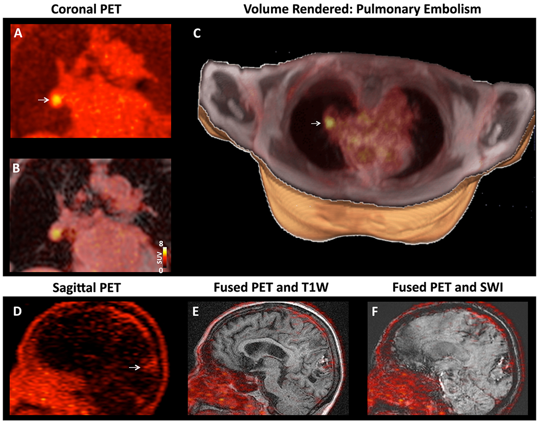
Figure 4. [64Cu]FBP8 facilitates whole-body thrombus detection.

(A-C) Right pulmonary artery embolism in a subject with a spontaneous LAA thrombus. (A) Coronal PET image with a focus of high [64Cu]FBP8 activity (arrow). (B) Fusion of the PET and Dixon water images reveals that the thrombus is located within a branch of the right pulmonary artery. (C) Volume rendered image showing the pulmonary embolism (arrow,) in the right lung. (D-F) TEE positive subject with a history of a large intracranial bleed. (D) Sagittal PET image, (E) fused PET and T1-weigted image and, (F) fused PET and susceptibility weighted image (SWI). The PET image shows that a low level of [64Cu]FBP8 uptake persists at the site of the bleed (arrow).
