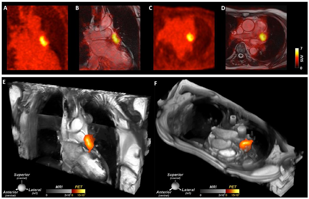
Central Illustration. Thrombus in the LAA induced by recent placement of a Watchman LAA closure device.

A large thrombus is seen in the LAA behind the device on (A, B) the coronal PET and PET/MRI images as well as (C, D) the axial PET and PET/MRI images. (E, F) 2D bSSFP cines stacks in the coronal and axial planes have been combined into a single 3D dataset and fused with the 3D PET data. In these multi-modal images the PET and MR data are both displayed in 3D, creating a volumetric depiction of the heart and the thrombus/[64Cu]FBP8 containing LAA. Volume rendered images in the oblique coronal and axial planes confirm the presence of thrombus in the LAA. (A-F) The thrombus produced by the closure device is contained within the LAA, and no evidence of thrombus is seen elsewhere in the heart or thorax.
