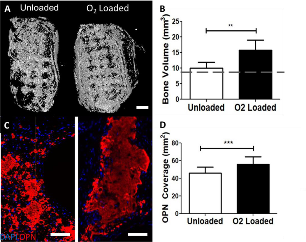
Figure 8: O2 delivery from μtank scaffolds increases mineralization and osteogenic protein deposition in ectopic defects.
Unloaded or O2-loaded 10% μtank scaffolds and ASCs were implanted into dorsal subcutaneous defects for 8 weeks. (A) CT of calvarial defects; Scale bar = 1 mm. (B) Quantification of bone area compared to empty defect; dashed line is average initial scaffold bone volume (n = 8). (C) Immunofluorescence images of osteopontin staining; scale bar = 100 μm. (D) Quantification of OPN immunostained area within the defects (n = 16–22). **, *** represent p < 0.01, 0.001).

