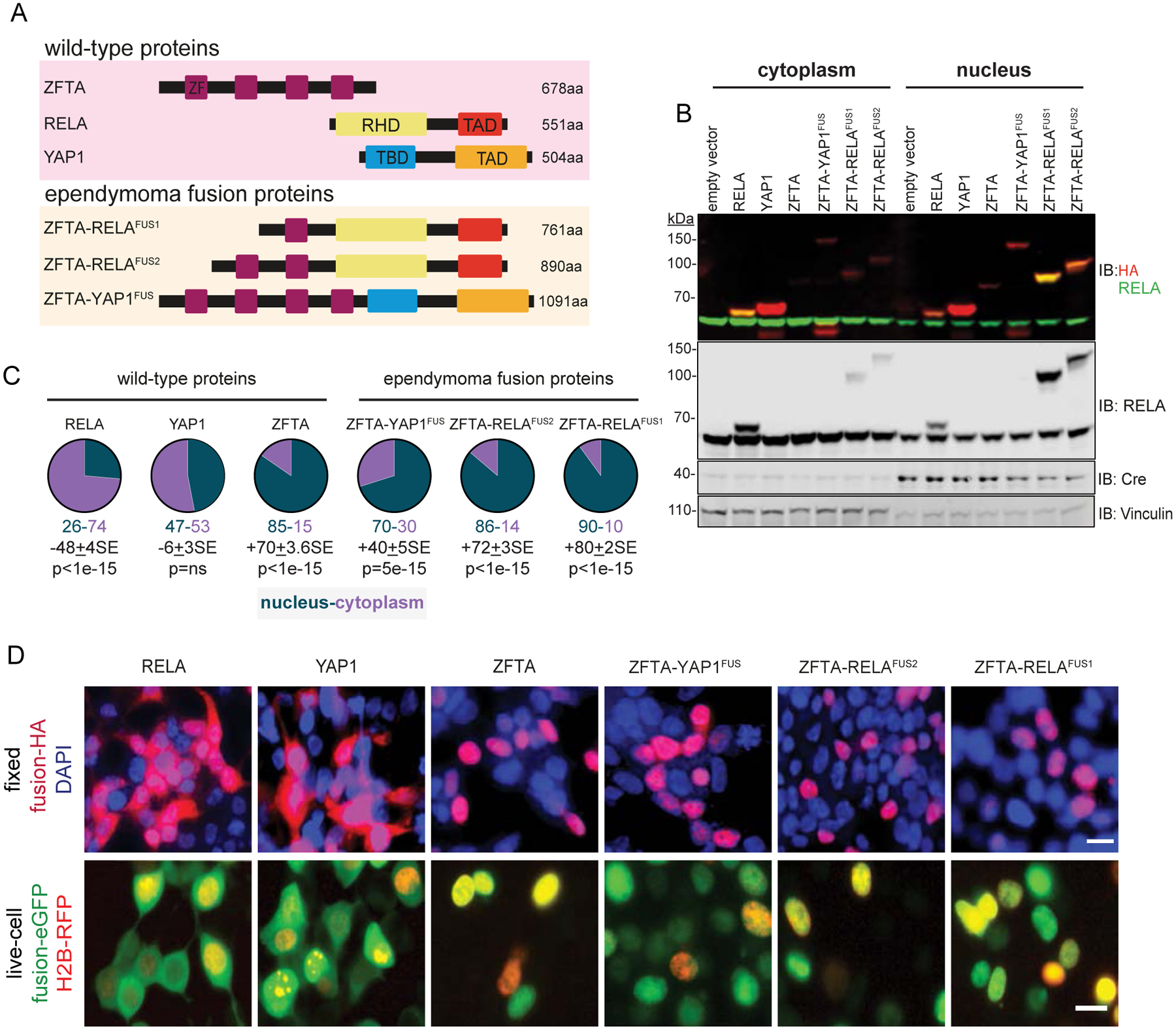
Figure 1. ZFTA-fusions translocate to the nucleus.

A. Schematics of wild-type and fusion proteins. Zinc Finger (ZF), REL homology (RHD), trans-activation (TAD) and TEAD-binding (TBD) domains. B. Immunoblots (IB) with indicated antibodies of cytoplasmic and nuclear fractions of HA-tagged proteins in HEK293 cells. Vinculin (cytoplasmic) and Cre (nuclear) are loading controls. C. Percent of proteins localized in cytoplasm or nucleus. Average difference + Standard Error (SE) and Mann-Whitney p-value (n=5), below. D. Top, localization of indicated HA-tagged proteins in DAPI counterstained and fixed HEK293 cells, or bottom, enhanced (e)-GFP tagged proteins in live, HEK293 cells expressing red fluorescence protein (RFP)-tagged Histone 2B (scale bar, 10μm).
