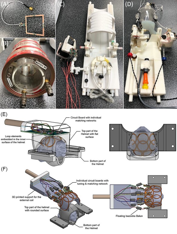
Figure 2 .

Radiofrequency coils used to image marmosets. (A) A surface coil, which allows for the flexibility of imaging across the marmoset body. (B) A commercially available volume coil, which also offers the flexibility of imaging across the marmoset body. Both (A) and (B) are less well suited for accelerated sequences (eg, fMRI). For this, custom-fitted phased array coils, such as the one shown in (C), which allows for imaging marmosets in stereotactic position under anesthesia, and (D-F), which are designed for imaging marmosets fully awake, are better suited. Panels (E) and (F) show the diagrams of an 8-element phased array coil embedded (E) or external (F) to the restraining helmet.
