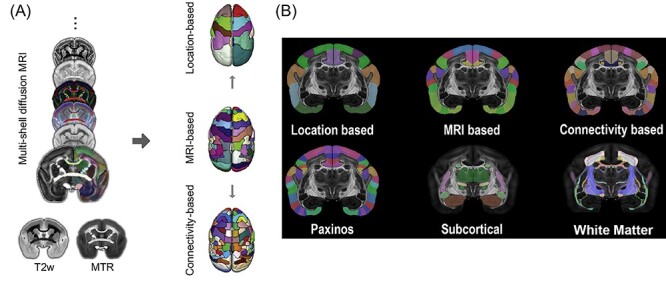
Figure 5 .

(A) The marmoset brain atlas was built from a set of ex-vivo multimodal high-resolution MRI images from the male marmoset brain sample that included multi-shell diffusion MRI, T2w, and magnetization transfer ratio (MTR). Based on the manifold local MRI contrasts, we manually delineated 54 cortical areas and 16 subcortical regions on 1 brain hemisphere. From this original version, we also created a coarser version with 13 larger cortical regions joined together based on their spatial locations, as well as a refined version in which 106 cortical areas were determined by performing a connectivity-based parcellation using diffusion tractography. (B) Top row: the marmoset brain parcellation based on the regional location (left), MRI segmentation (middle), and connectivity-based analysis (right). Bottom-row: the marmoset brain parcellation based on the Paxinos atlas (left), subcortical structures (middle), and white-matter fiber pathways (right).
