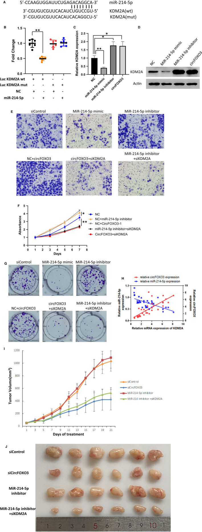
FIGURE 4.

circFOXO3 up‐regulates KDM2A expression by targeting miR‐214. A, Binding sites between miR‐214 and KDM2A was shown. B, Luciferase activity was measured in miR‐214 and KDM2A WT, Mut cDNA plasmids infected 293T. Data are presented as means ± SD (n = 3). **P < .01. C, Relative expression of KDM2A in SCC‐4 cells with indicated treatment. Data are presented as means ± SD (n = 3). *P < .05, **P < .01. D, KDM2A protein level was measured by western blot of SCC‐4 with indicated treatment. E, The effects of KDM2A on both SCC‐4 cell invasion. A total of 1 × 10⁵ transfected cells were seeded onto a permeable membrane in a Boyden chamber to allow the cells to invade the opposite layer of the membrane. F, SCC‐4 cells were transfected as indicated and cultured for different times. Cell viability was measured by CCK‐8 assay. Data represent the means ± SD (n = 3). *P < .05, **P < .01. G, Proliferation of SCC‐4 cells transfected with indicated siRNA/plasmid, as detected by colony formation assay. H, The correlation of miR‐214/KDM2A, circFOXO3/KDM2A expression level in OSCC tumor tissues analyzed by Pearson. I, Four‐week‐old nude mice were engrafted with 1 × 10⁷ SCC‐4 cells and randomly divided into four groups (n = 6). After two weeks, the tumor‐bearing mice were treated with adenovirus expressing different siRNA or inhibitor as indicated. Tumor volumes were calculated by measuring the length and width using Vernier calipers every two days. Data represent the means ± SD (n = 6). J, Images of the tumors for Figure 4I
