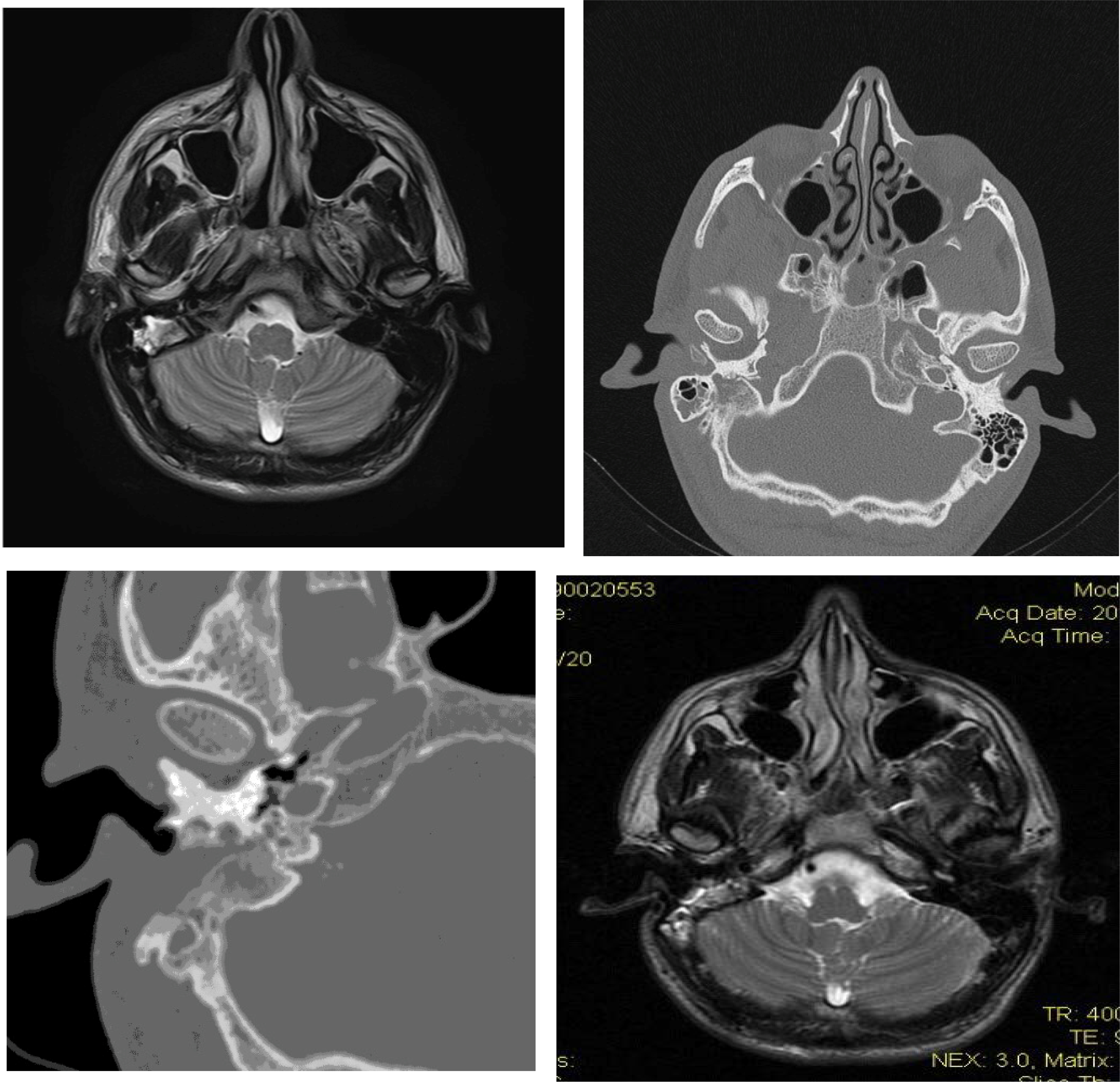
FIGURE 1.

a: Axial MRI of visceral cranium: Solid-tissue mass of the right temporal bone with hyperintense pathological magnetic signal and peripheral areas of enhancement after gadolinium administration
b,c: CT of the temporal bone. An invasive soft tissue density lesion arising from the right temporal bone occupying partially the right mastoid cavity-with corrosion of the trabeculae of bone
d: Axial MRI of visceral cranium (2 years later): Hyperintense signal in the resection cavity with clear reduction of the pathological signal and of the intake of gadolinium in the right temporal bone
