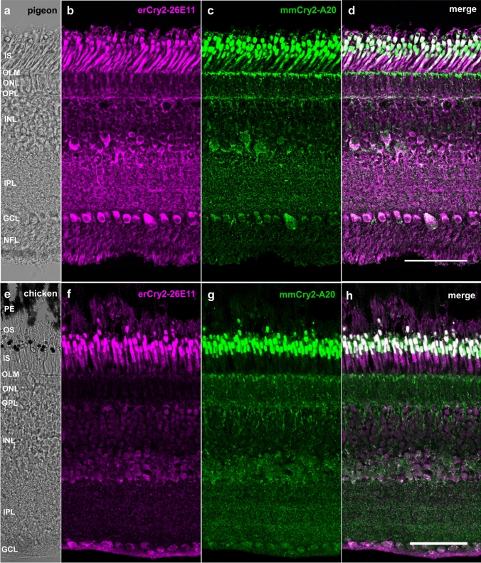
Fig. 3.
Cry2 location was confirmed in two additional bird species. The antibodies erCry2-26E11 and mmCry2-A20 stained the photoreceptor inner segments as well as the cytoplasm of cells in the inner nuclear layer and ganglion cell layer in the day-flying pigeon (a–d) and the domestic chicken (e–h). mmCry2-A20 was also labelling the outer limiting membrane in the pigeon (c) and the chicken (g). In contrast to the immunosignals observed in the European robin retina, mmCry2-A20 did not label the nuclei but the cytoplasm in pigeon and chicken. Images a and e are bright-field images, images b–d and f–g are maximum projections of confocal stacks (b, c: z-size 4 µm, 20 sections; f–h: z-size 2 µm, 10 sections). Scale bars: d, h 50 µm. PE retinal pigment epithelium, OS photoreceptor outer segments, IS photoreceptor inner segments, OLM outer limiting membrane, ONL outer nuclear layer, OPL outer plexiform layer, INL inner nuclear layer, IPL inner plexiform layer, GCL ganglion cell layer, NFL nerve fibre layer

