Fig. 5.
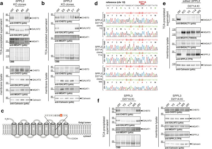
Validation of novel SPPL3 substrates in SPPL3 KO and SPPL3 D271A knock-in cells. a Immunoblot validation of the newly identified SPPL3 substrates CHST3, GALNT2 and MGAT1 in HEK293 cells. TCA-precipitated CM and membrane lysates obtained from three distinct SPPL3 KO clones and parental HEK293 cells (ctrl) were probed using specific antibodies. Calnexin served as loading control for the membrane fraction, total protein staining with Coomassie Brilliant Blue (CBB) as loading control for CM samples. b Immunoblot validation of the CHST3, GALNT2 and MGAT1 in HeLa cells. c Overview of SPPL3 topology (loops are not to scale). D271 targeted is highlighted in red, the GIGD motif in bold font. d Sanger sequencing reads confirming correct genome editing in SPPL3 D271A knock-in HEK293 cells. The targeted region of the SPPL3 locus was amplified by PCR from genomic DNA isolated from the indicated clones, gel-purified and Sanger sequenced. Reads were aligned to the corresponding chromosome 12 reference sequence. Encoded SPPL3 primary structure is provided for the correct reading frame. Mutated residues are highlighted by a red background. All clones analysed carry only the desired D271A missense mutation (red aa residue) in the active site of SPPL3. Additional mutations were introduced to prevent Cas9 re-cleavage, but remained silent (orange aa residues). e Phenotypic validation of SPPL3 D271A knock-in cells in comparison to SPPL3 KO and parental HEK293 cells. Secretion of B4GALT1 from cells was examined by immunoblotting of TCA-precipitated samples. Lysates of carbonate-washed membranes from the indicated cell clones were probed for the SPPL3 substrate MGAT5 and B4GALT1 as well as for SPPL3. f Immunoblot analysis of CHST3, GALNT2 and MGAT1 levels in TCA-precipitated supernatant and membrane lysates of isogenic SPPL3 D271A HEK293 cells

