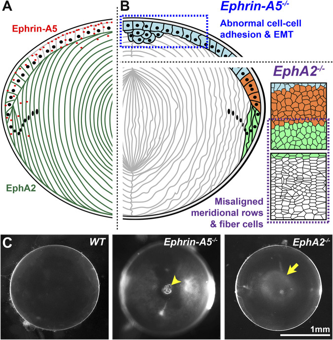
FIGURE 3.
EphA2 and ephrin-A5 in mouse lenses. (A) EphA2 (green) is mainly expressed in equatorial epithelial cells and lens fiber cells, while ephrin-A5 (red) is mainly present in anterior epithelial cells with some expression in peripheral fiber cells and in fiber cell tips near the lens suture. (B) In C57BL/6J genetic background mice, loss of ephrin-A5 leads to abnormal cell-cell adhesion between anterior epithelial cells and epithelial-to-mesenchymal transition (EMT) of these normally quiescent cells. In contrast, disruption of EphA2 in C57BL/6J mice leads to disorder of the equatorial epithelial cells, which leads to abnormal lens fiber cell shape. (C) The normal wild-type (WT) lens is clear on a darkfield background. In contrast, ephrin-A5 −/− lenses often have anterior cataracts (arrowhead), and EphA2 −/− lenses often display nuclear cataracts at the center of the lens (arrow). These images are of lenses from three-week-old mice in the C57BL/6J genetic background. Modified from (Cheng et al., 2017). Illustrations are not drawn to scale. Scale bar, 1 mm.

