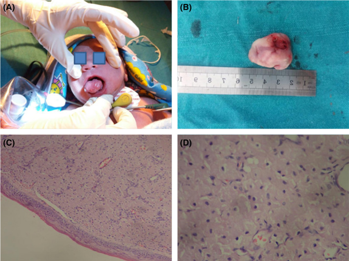
FIGURE 1.

(A) Examination of the case of congenital granular cell tumor (CGCT), (B) Excised specimen of CGCT in one‐day‐old neonate, (C) Granular cells in a fibrovascular stroma, lined by thin, atrophic epithelium (H&E‐40× magnification), (D) Large, round granular cells with basophilic nuclei and abundant eosinophilic cytoplasm (H&E‐100× magnification)
