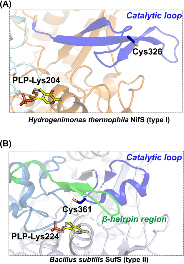
Figure 5 .

(A) Active site structure of Hydrogenimonas thermophila NifS [PDB ID: 5zsp]. (B) Active site structure of Bacillus subtilis SufS [PDB ID: 5zs9]. Catalytic loops in both NifS and SufS are colored in blue. The β-hairpin region of SufS is colored in green. The PLP-Lys moieties and catalytic cysteine residues are shown in stick models.
