
Figure 6 .
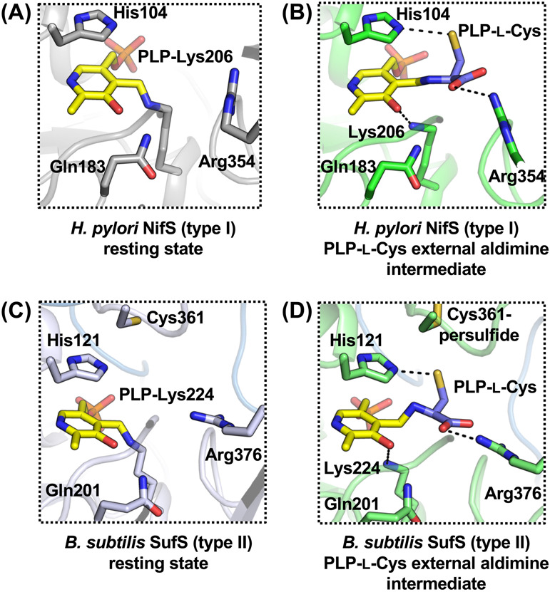
(A) Active site structure of Helicobacter pylori NifS in the resting state [PDB ID: 5wt2]. (B) Active site structure of H. pylori NifS in an intermediate state of PLP-l-Cys external aldimine [PDB ID: 6kg0]. (C) Active site structure of Bacillus subtilis SufS in the resting state [PDB ID: 5zs9]. (D) Active site structure of B. subtilis SufS in an intermediate state of PLP-l-Cys external aldimine with Cys361-persulfide [PDB ID: 6kfz]. Dashed lines indicate key polar interactions for binding of the substrate l-cysteine into the active sites.