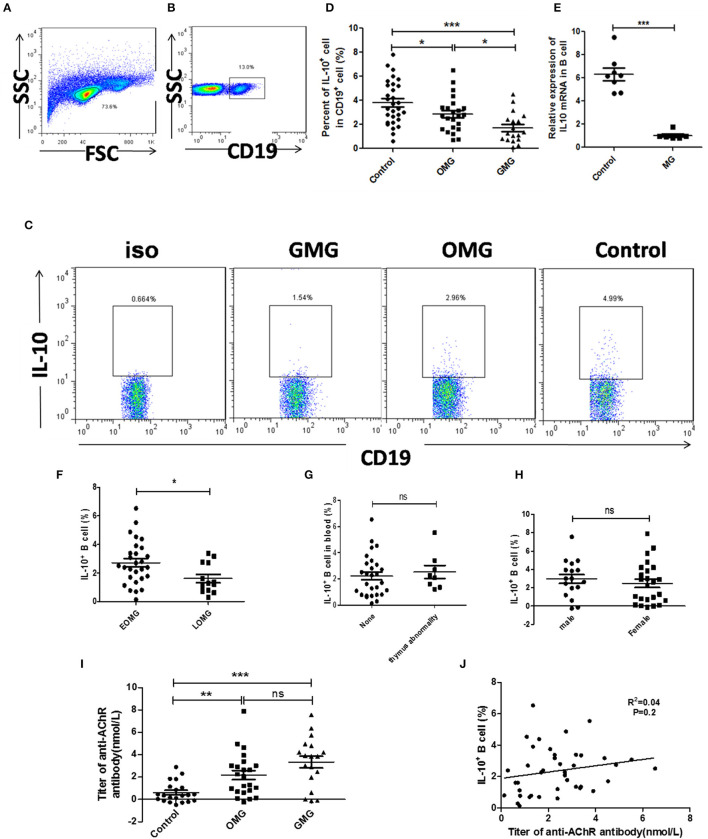
Figure 1.
The proportion of CD19+ IL-10+ cells in peripheral blood cells of MG patients and controls. (A) Lymphocytes were gated according to forward scatter and side scatter by FCM. (B) B cells were gated according to CD19+ in lymphocytes. (D) Scatter plots show the mean percentages of CD19+ IL-10+ cells in the peripheral blood of 23 OMG patients, 18 GMG patients, and 30 healthy individuals. (E) Expression of IL-10 mRNA by real-time quantitative analysis in the peripheral blood of 8 MG patients and 8 healthy individuals. Six of 8 MG patients were ocular myasthenia gravis. (C) Representative flow cytometry plot of CD19+ IL-10+ cell gating for patients with OMG, GMG, and healthy controls. (F) The frequency of CD19+ IL-10+ cell in different MG types according to age at onset. (G) The frequency of CD19+ IL-10+ cells in different thymus status. (H) The frequency of CD19+ IL-10+ cells in different gender. (I) The titer of anti-AChR antibody in MG patients, including 21 healthy controls, 23 OMG patients, and 18 GMG patients. (J) The proportion of CD19+ IL-10+ cells had no correlation with the level of anti-AChR antibody, and the samples were from 41 patients with MG. p-values were calculated by Student's t-test; *p < 0.05, **p < 0.01, ***p < 0.001, ns, not significant.

