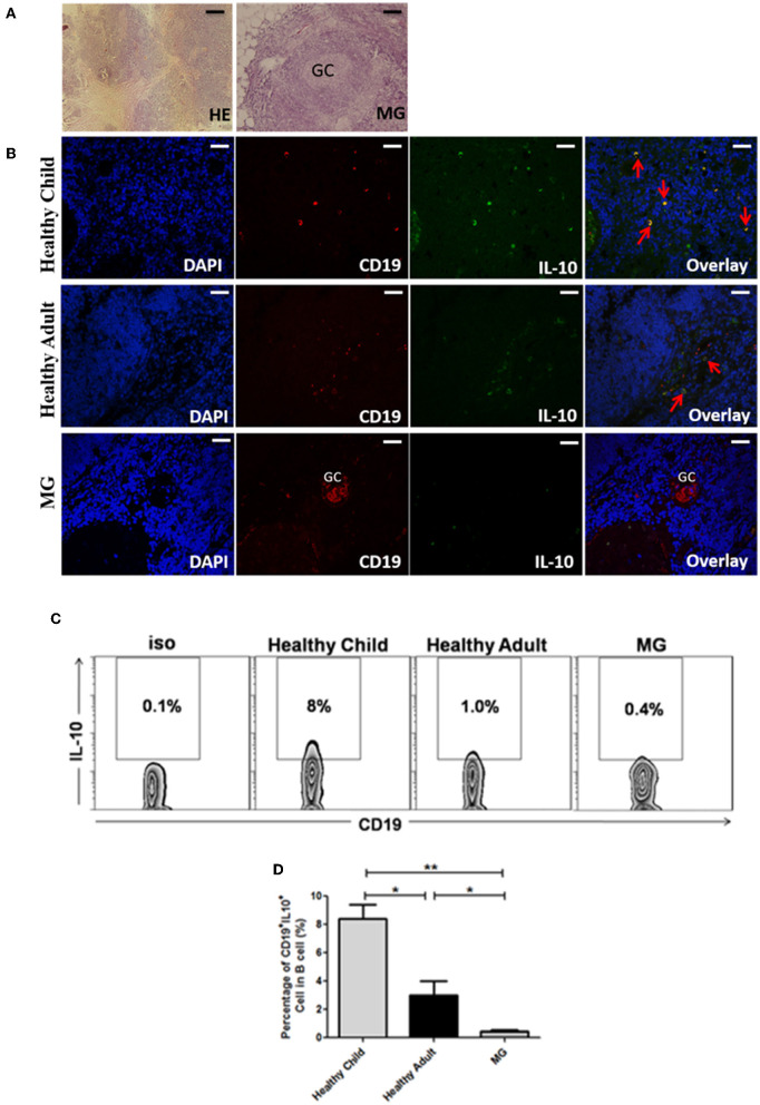
Figure 4.
CD19+IL-10+ Breg cells were assessed in the thymus of healthy individuals and MG patients by immunofluorescence staining. (A) Human normal thymus and hyperplastic thymus from MG patients stained with hematoxylin and eosin. In addition, thymic hyperplasia was observed in an MG patient, which showed a germinal center (GC) structure (scale bars, 100 μm). (B) Frozen sections of thymic tissues were immunoassayed with nuclear DAPI staining (blue). Double immunofluorescence analysis was performed with anti-CD19 (red) and anti-IL-10 (green) antibodies. Note that CD19+IL-10+ Breg cells both express CD19 and IL-10 (yellow, with red arrow). It showed a germinal center (GC) structure in the hyperplastic thymus from MG patient (scale bars, 200 μm). Immunofluorescence control showed negative (not shown). (C) Representative dot plot showing the percentage of CD19+IL-10+ Breg cells in CD19+ B cells in the thymus of healthy children, healthy adults, and MG patients. (D) Bar charts showing the mean percentage ± SE of CD19+IL-10+ Breg cells in CD19+ B cells in the thymuses of healthy children (n = 10), healthy adults (n = 4), and MG patients (n = 13, 5 of MG thymuses are thymoma and 7 of MG thymuses are thymic hyperplasia); *p < 0.05, **p < 0.01.

