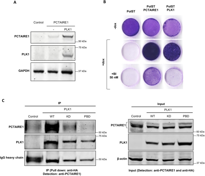
Fig. 6.
PCTAIRE1 interacts with PLK1. (A) After transient transfections of 1 µg each of FLAG-PCTAIRE1 and myc-PLK1 in 293T cells, PCTAIRE1 was stabilized. (B) Crystal Violet staining of U2OS-PolST cells showing reversal of cell survival in the presence of PLK1 inhibitor BI-2536 (50 nM) in PLK1-overexpressing PolST (PolST-PLK1) cells but not in PCTAIRE1-overexpressing PolST (PolST-PCTAIRE1) cells. (C) HA-tagged constructs of wild-type PLK1 (WT), PLK1 kinase-dead mutant (KD) and PLK1 polo-box domain mutant (PBD) were transiently transfected in U2OS cells (1 µg each). After pulling down PLK1 by anti-HA antibody, PCTAIRE1 was co-immunoprecipitated with WT-PLK1 but not with KD and PBD mutants.

