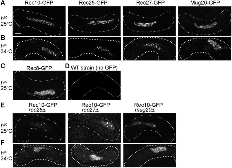
Fig. 1.
LinE proteins form elongated linear structures in live zygotic meiosis. (A-F) Rec10, Rec25, Rec27, Mug20 and Rec8 were observed as GFP fusion proteins in h90 (homothallic) strains using SIM. Images are maximal projections of the z-stack (see Fig. S1). The dotted line represents the cell outline. Each image is representative of ≥20 horsetail stage cells (i.e. meiotic prophase; Ding et al., 2004). (A) All LinE proteins form elongated structures in zygotic meiosis at 25°C. (B) Rec10, Rec27 and Rec25 fail to form linear structures at high temperature (34°C); instead, they form non-continuous structures and often bright nuclear dots. These ‘dotty foci’ differ from the ‘linear structures’ at 25°C (Fig. 1A) but are like previously reported structures in azygotic meiosis at 34°C (Davis et al., 2008; Estreicher et al., 2012; Fowler et al., 2013). Mug20 shows long linear structures at 34°C, like those at 25°C (Fig. 1A; see also Ding et al., 2021). (C) Rec8 forms nearly end-to-end chromosomal structures in zygotic meiosis at 25°C (Ding et al., 2016). (D) Wild-type (WT, no GFP) h90 cells show limited green fluorescence in zygotic meiosis at 25°C. (E) At 25°C in zygotic meiosis of LinE deletion mutants, Rec10 forms a few bright nuclear foci and more nearly uniform nuclear distribution than in wild type (Fig. 1A). Thus, Rec10 still enters the nucleus and forms a few nuclear foci, but not linear structures, in the absence of any other LinE protein. (F) At 34°C in zygotic meiosis of LinE deletion mutants, Rec10 forms a few nuclear foci and nearly uniform nuclear distribution, more prominent than at 25°C. Scale bar: 2 μm.

