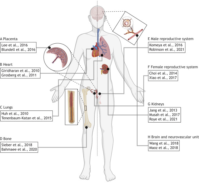
Fig. 6.
Microfluidic devices for modeling the development and function of various tissues and organs. (A) Microfluidic devices have been used to model the placental barrier (Lee et al., 2016) and have been used to recapitulate maternal-fetal crosstalk (Blundell et al., 2016). (B) Contractile myocytes are commonly used to produce cardiac microfluidic devices (Grosberg et al., 2011) and these systems can subsequently be used to model pharmacological responses (Giridharan et al., 2010). (C) The lungs were one of the first organs modeled using microfluidic devices (Huh et al., 2010), and processes involved in fetal lung development have also been observed (Tenenbaum-Katan et al., 2015). (D) Cells cultured in a microfluidic device mimicking the bone marrow niche are capable of differentiating into specialized types (Sieber et al., 2018), including osteoblasts which facilitate osteogenesis (Bahmaee et al., 2020). (E) Testicular tissue explants cultured in microfluidic devices can generate functional spermatozoa (Komeya et al., 2016), and bioprinted testicular cells organize into their native cytoarchitecture while maintaining their functionality (Robinson et al., 2021 preprint). (F) The role of mechanical heterogeneity in the ovulation of ovarian follicle explants has been studied using microfluidic devices (Choi et al., 2014), and the relationship between the female reproductive tract and the endocrine system has also been studied using connected microfluidic modules (Xiao et al., 2017). (G) The absorption and filtration functions of the kidney proximal tubules (Jang et al., 2013) and glomerulus (Musah et al., 2017; Roye et al., 2021), respectively, have been modeled using microfluidic devices. (H) Microfluidic devices facilitate the differentiation and organization of brain organoids for studying developmental diseases or pharmacological toxicity (Wang et al., 2018). Similarly, multiple microfluidic devices can be coupled to model the blood-brain barrier for observing cell crosstalk and barrier function (Maoz et al., 2018). Anatomical diagram was created with BioRender.com.

